Fusarium keratitis is a filamentary fungal corneal infection that can threaten vision within weeks.
It often follows plant trauma in warm agricultural regions, although contact lens wear also carries risk.
For the general approach, see fungal keratitis diagnosis and treatment.
Risk Factors for Fusarium Keratitis
Corneal trauma with vegetable matter, soil or wood is the classic history.
Farmers and outdoor workers form a familiar high-risk group.
Prior topical corticosteroids, ocular surface disease and corneal surgery also favor fungal infection.
However, some patients recall no injury.
Symptoms Patients Report
Patients usually describe pain, redness, tearing, photophobia and blurred vision.
Many arrive after failed antibiotic drops.
For example, a farmer with a worsening ulcer after a week of fluoroquinolones deserves fungal testing.
All-fit Slit-Lamp Adapter
Record and share exactly what you see at the slit lamp. One adapter fits any slit lamp or surgical microscope — and any smartphone.
From Choroida — the team behind this siteContact Lens Wear
In 2006, a multistate outbreak linked Fusarium keratitis to one contact lens solution.
Therefore, ask every lens wearer about solutions, storage and overnight wear (see contact lens risk factors).
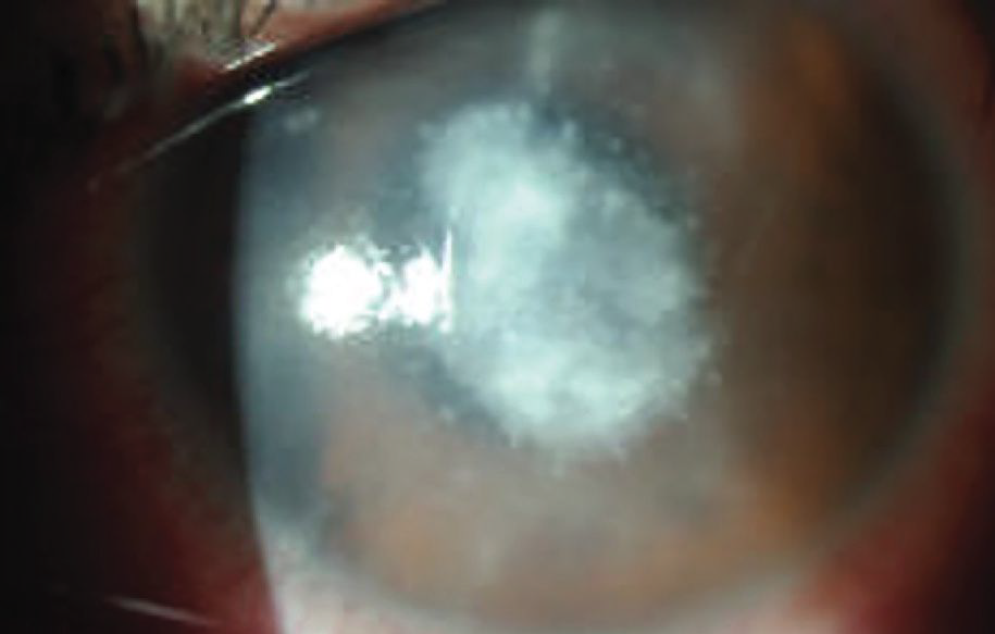
Feathery Infiltrate in Fusarium Keratitis
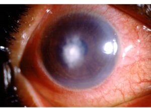
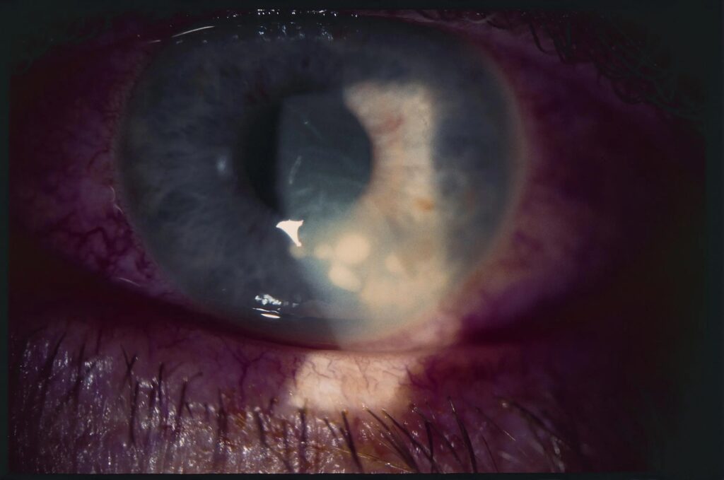
At the slit lamp, the infiltrate looks gray-white, dry and slightly raised.
Its margins appear feathery because hyphae extend along the stromal lamellae.
Satellite lesions, an immune ring, an endothelial plaque or hypopyon may also appear.
Examination Pearls in Fusarium Keratitis
First, measure the infiltrate and epithelial defect with the slit lamp beam and record both dimensions.
Then check for an endothelial plaque, anterior chamber reaction and intraocular pressure.
Also photograph the lesion, because serial images show trends more reliably than memory.
Therefore, compare each visit against the same baseline.
Deep Invasion
Hyphae can penetrate deep stroma and reach Descemet membrane.
As a result, surface scrapings may turn negative because the organism sits below the sampled tissue.
Anterior segment OCT documents depth and thinning at each visit.
Differential Diagnosis
Bacterial keratitis is the main mimic, especially after partial treatment (see fungal versus bacterial clues).
Also consider Acanthamoeba, Nocardia and herpetic stromal disease.
Confirming Fusarium Keratitis in the Lab
Scrape the ulcer base and leading edge before starting antifungal therapy.
Next, send material for KOH or calcofluor white microscopy and for fungal cultures.
If scrapings stay negative, a corneal biopsy can sample deeper tissue.
Confocal microscopy can show branching hyphae, but it cannot name the genus.
Natamycin for Fusarium Keratitis
Natamycin 5% suspension is the usual first-line drug.
It binds ergosterol in the fungal cell membrane.
Dosing is often hourly while awake, then tapers according to response.
However, penetration through intact epithelium is limited, so gentle debridement may help.
Natamycin can also cause surface toxicity that mimics slow healing.
What MUTT I Showed
The Mycotic Ulcer Treatment Trial I compared topical natamycin with topical voriconazole.
Natamycin produced better visual acuity and fewer perforations or therapeutic grafts.
Moreover, the benefit appeared concentrated in Fusarium ulcers.
Because the trial enrolled patients in South India, local isolates may behave differently elsewhere.
Voriconazole in Fusarium Keratitis
MUTT II tested oral voriconazole added to topical therapy and found no overall benefit.
A subgroup analysis suggested possible benefit for Fusarium, although this needs confirmation.
Meanwhile, oral voriconazole can cause liver enzyme elevation and visual disturbances.
Therefore, discuss these risks and arrange monitoring when specialists choose systemic treatment.
Drug Susceptibility Testing
In MUTT I isolates, higher minimum inhibitory concentrations were associated with poorer outcomes.
Testing can therefore add information when an ulcer fails to improve.
However, laboratories vary, and results cannot replace serial examination.
Adjuncts and Their Limits
Intrastromal voriconazole showed no benefit in a randomized trial of severe fungal ulcers.
Similarly, adjuvant cross-linking did not improve outcomes in the CLAIR trial.
Finally, stop topical corticosteroids during active infection, because they can promote fungal growth.
Judging Response in Fusarium Keratitis
Fungal ulcers improve slowly, so expect gradual change over days to weeks.
Encouraging signs include blunted margins, a smaller infiltrate and epithelial healing.
Conversely, expanding infiltrate, progressive thinning or new endothelial plaque suggests failure.
Surgery for Fusarium Keratitis
Therapeutic keratoplasty is an option for perforation, limbal spread or progression despite maximal therapy.
However, reinfection and graft failure remain risks, so surgery cannot guarantee useful vision.
Follow-Up After Healing
After the epithelium closes, however, residual stromal scarring may still limit vision.
Therefore, taper antifungal drops slowly and keep watching for relapse.
Counseling Patients
Explain that treatment is intensive and may last several weeks.
Patients should also return early for increasing pain or discharge.
Next, discuss scarring, possible surgery and strict lens avoidance.
Pitfalls to Avoid
- Do not treat a post-trauma ulcer as bacterial without cultures
- Avoid steroids before excluding fungal infection
- Do not rely on a negative superficial scrape in a deep lesion


Document what you see
Two smartphone imaging tools built for everyday clinic use — one for the slit lamp, one for the fundus.
From Choroida — the team behind this siteReferences
- Prajna NV, Krishnan T, Mascarenhas J, et al. The Mycotic Ulcer Treatment Trial: a randomized trial comparing natamycin vs voriconazole. JAMA Ophthalmol. 2013;131:422-429.
- Prajna NV, Krishnan T, Rajaraman R, et al. Effect of oral voriconazole on fungal keratitis in the Mycotic Ulcer Treatment Trial II (MUTT II): a randomized clinical trial. JAMA Ophthalmol. 2016;134:1365-1372.
- Chang DC, Grant GB, O’Donnell K, et al. Multistate outbreak of Fusarium keratitis associated with use of a contact lens solution. JAMA. 2006;296:953-963.
- Prajna NV, Radhakrishnan N, Lalitha P, et al. Cross-Linking-Assisted Infection Reduction: a randomized clinical trial evaluating the effect of adjuvant cross-linking on outcomes in fungal keratitis. Ophthalmology. 2020;127:159-166.

Writes and reviews the clinical articles on Educate. Every post is checked against peer-reviewed sources before it goes live.