
Uveal melanoma prognostic testing estimates the risk that a tumor will metastasize, using molecular features of the tumor cells.
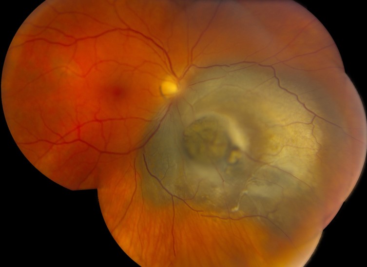
It does not change the treatment of the eye, but it informs surveillance and counselling.
Because there is no proven treatment to prevent metastasis, experts debate the benefits and the ethics of testing.
Why Uveal Melanoma Prognostic Testing Is Done
About half of patients with uveal melanoma eventually develop metastases, most to the liver.
The interval can be many years.
Clinical features such as tumor size, ciliary body involvement, and extraocular extension predict risk, but they are imprecise (see choroidal melanoma).
Molecular tests identify tumors with high-risk biology.
They can also distinguish patients who need close monitoring from those at lower risk.
Uveal Melanoma Prognostic Testing: Cytogenetic Features
- Monosomy of chromosome 3, the best-known marker, strongly associated with metastasis
- Gain of chromosome 8q, also associated with a worse outlook, especially with monosomy 3
- Gain of chromosome 6p, associated with a better outlook
Laboratories can test chromosomes on tumor tissue obtained by fine-needle biopsy or after enucleation.
Fundus Explorer Pro
Photograph the retinal findings described here with the phone already in your pocket — 22 D optics and built-in illumination in one handheld unit.
From Choroida — the team behind this siteGene Expression Profiling
Onken and colleagues described two molecular classes of uveal melanoma on the basis of gene expression: class 1, with low metastatic risk, and class 2, with high risk.
A commercial assay measures the expression of a panel of genes and assigns the tumor to class 1A, 1B, or 2, with a corresponding risk of metastasis.
A prospective multicenter study validated the test, showing that class 1A tumors had a low rate of metastasis at five years, and class 2 tumors had a high rate (see the references).
Uveal Melanoma Prognostic Testing: Mutational Markers
- BAP1 mutations, found in a large share of metastasizing tumors, linked to loss of one copy of chromosome 3 and to a high risk
- SF3B1 and EIF1AX mutations, associated with intermediate and low risk respectively
- GNAQ and GNA11, early driver mutations that do not predict outcome
- PRAME expression, an additional marker used with gene expression profiling in some assays
Harbour and colleagues found frequent BAP1 mutations in metastasizing uveal melanomas, which helped to explain the biology.
How the Sample Is Obtained
- Fine-needle aspiration biopsy of the tumor, usually at the time of plaque placement or in an office setting
- Enucleation specimen from a removed eye
- Transscleral or transvitreal approaches, according to the tumor location
Biopsy carries small risks, including hemorrhage, retinal detachment, and tumor seeding, and the sample may be too small for testing.
Sampling error is a concern, since tumors are heterogeneous, and a normal result may not represent the whole tumor.
What the Result Changes
- Surveillance intensity: patients with high-risk tumors usually have liver imaging, by ultrasound, MRI, or CT, and examination at intervals of three to six months, while low-risk patients can be followed less often
- Counselling: it gives patients and families a realistic estimate of the risk
- Clinical trials: high-risk patients may also be eligible for adjuvant trials
- Reassurance: a class 1A result may also spare a patient anxiety and over-testing
Currently, no adjuvant therapy has been proven to improve survival, so a high-risk result does not lead to a proven treatment.
Uveal Melanoma Prognostic Testing: Limitations and Ethics
- Not all patients want to know their risk, so offer testing after counselling
- Results can cause distress, particularly when no preventive treatment exists
- The test has limited value for small, low-risk tumors, where the chance of metastasis is already small
- Costs and availability vary between centers
Testing is best done at a center with experience.
The results should be discussed with the patient by a team that includes an ocular oncologist.
Practical Advice
Document the clinical features and the testing results in the chart, and arrange regular surveillance with a clear schedule.
Also, provide the patient with information about the disease and support resources.
Communicating the Result
Discuss the result with the patient in terms of probabilities and what they mean for follow-up, and not as a prediction of fate.
Also, allow time for questions and for the patient to bring a family member, and provide information from a reliable organization.
Offer psychological support to those who are also distressed, and remind them that the test describes a risk and that many patients with high-risk features live for years.
In addition, make sure that the patient knows who will coordinate the surveillance imaging and how the team will communicate results.
For the latest studies, see the PubMed results for uveal melanoma prognostic testing.


Document what you see
Two smartphone imaging tools built for everyday clinic use — one for the slit lamp, one for the fundus.
From Choroida — the team behind this siteReferences
- Onken MD, Worley LA, Ehlers JP, Harbour JW. Gene expression profiling in uveal melanoma reveals two molecular classes and predicts metastatic death. Cancer Res. 2004;64:7205-7209.
- Onken MD, Worley LA, Char DH, et al. Collaborative Ocular Oncology Group report number 1: prospective validation of a multi-gene prognostic assay in uveal melanoma. Ophthalmology. 2012;119:1596-1603.
- Harbour JW, Onken MD, Roberson EDO, et al. Frequent mutation of BAP1 in metastasizing uveal melanomas. Science. 2010;330:1410-1413.
- American Academy of Ophthalmology. Basic and Clinical Science Course, Section 4: Ophthalmic Pathology and Intraocular Tumors. AAO; current edition.

Writes and reviews the clinical articles on Educate. Every post is checked against peer-reviewed sources before it goes live.


