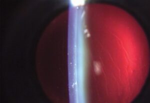
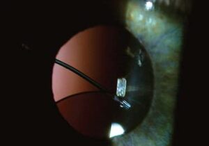
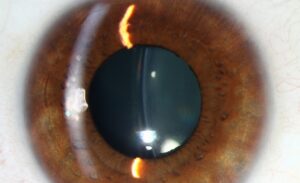
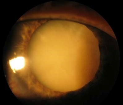
A brunescent cataract is an advanced nuclear cataract in which the nucleus has changed from yellow to deep brown, and, in the most extreme cases, to almost black.
The name cataracta nigra is used for this last stage.
These lenses are hard, and they are the ones that surgeons approach with the most caution.
Why the nucleus darkens
Throughout life, the nucleus of the lens becomes denser and more compact, as new fibers are laid down around the old ones.
Proteins in the nucleus aggregate and are modified by oxidation, glycation, and ultraviolet exposure, and they become cross-linked and pigmented.
The progression is from clear, to yellow, to amber, to brown, and finally to black.
Patients who have lived in tropical, high-altitude and high-UV environments, those who smoke, and those with diabetes and long-standing steroid use develop brunescence earlier.
In places where cataract surgery is delayed for economic or access reasons, brunescent cataracts are seen much more often than in places with early surgery.
What the patient experiences
- Vision loss that can be marked, with reduced contrast and color perception, and a perception that everything has a brown tinge
- Improved near vision for a time in some patients, due to the myopic shift (“second sight”), which gradually lessens as the nucleus darkens
- Glare, particularly in the dark
- In extreme cases, only light perception
On examination, the red reflex is dull or absent, the fundus is hard to see, and the lens may be seen as a dark brown mass behind the pupil.
Pseudophakic comparison of the two eyes may reveal a marked difference.
All-fit Slit-Lamp Adapter
Record and share exactly what you see at the slit lamp. One adapter fits any slit lamp or surgical microscope — and any smartphone.
From Choroida — the team behind this siteWhy surgery is more difficult
- The nucleus is hard and needs more ultrasound energy to emulsify, which increases heat and turbulence and puts the corneal endothelium at risk (see corneal endothelial decompensation)
- Hard nuclei are heavy, and fragments can damage the posterior capsule or fall into the vitreous
- Zonular stress is greater, particularly in patients with pseudoexfoliation or trauma
- Poor red reflex makes capsulorhexis harder, and trypan blue staining of the anterior capsule is often necessary
- The risk of posterior capsule rupture and vitreous loss is higher (see vitreous loss in cataract surgery)
- Wound burns and corneal edema are more frequent postoperatively
How surgeons plan for it
- Preoperative specular microscopy to assess endothelial reserve
- Use of dispersive and cohesive viscoelastic together (the soft shell technique) to protect the endothelium
- Phaco chop or divide-and-conquer techniques that reduce the energy used
- Careful hydrodissection and nuclear rotation
- Staining the capsule
- Considering manual small incision cataract surgery or extracapsular extraction when the nucleus is very dense and the endothelium is poor, particularly where phaco resources are limited
Postoperative course
Corneal edema is common in the first days and usually clears.
Dense nuclear fragments left behind or an IOP spike should be checked for.
Patients should be told about the possibility of prolonged edema, and about the longer recovery time compared with softer cataracts.
Talking to the patient before surgery
Patients with dense cataracts need honest information. Explain that the operation is more demanding, that the cornea may take longer to clear, that the risk of complications is higher than for a soft cataract, and that the final vision depends on the retina and optic nerve, which cannot be examined directly. Reassure them that, with good planning, most do very well, and that delaying further will only make the lens harder.
Assessing the back of the eye
Dense lenses hide the fundus, so a B-scan ultrasound is essential to exclude a detachment, a tumor, or a staphyloma. Check the pupil for a relative afferent defect, since an unexpected defect suggests optic nerve or retinal disease. Light projection and color perception are simple clinical tests that give some information about retinal function.
Early surgery as prevention
Public eye care programs have shown that earlier surgery reduces the number of brunescent lenses and their complications. In clinical practice, a patient who has had a gradual decline of vision and refuses surgery should be informed that the cataract will not shrink, and that waiting may make the operation less safe.
Prognosis
Visual results can be excellent when the retina and optic nerve are healthy, but the risk of complications is higher.
The best strategy is early surgery, before the nucleus becomes so hard.


Document what you see
Two smartphone imaging tools built for everyday clinic use — one for the slit lamp, one for the fundus.
From Choroida — the team behind this siteReferences
- Kelman CD. Phaco-emulsification and aspiration: a new technique of cataract removal. Am J Ophthalmol. 1967;64:23-35.
- Ruit S, Tabin G, Chang D, et al. A prospective randomized clinical trial of phacoemulsification vs manual sutureless small-incision extracapsular cataract surgery in Nepal. Am J Ophthalmol. 2007;143:32-38.
- Chylack LT Jr, Leske MC, McCarthy D, et al. Lens opacities classification system II (LOCS II). Arch Ophthalmol. 1989;107:991-997.
- Gogate P, Optom JJ, Deshpande S, Naidoo K. Meta-analysis to compare the safety and efficacy of manual small incision cataract surgery and phacoemulsification. Middle East Afr J Ophthalmol. 2015;22:362-369.
Test yourself
A few questions straight from this article.
-
What does the term cataracta nigra describe?
Cataracta nigra is the name given to the most extreme stage of a brunescent cataract, in which the deep brown nucleus has become almost black. -
In what order does the lens nucleus change colour as a brunescent cataract develops?
The nucleus progresses from clear, to yellow, to amber, to brown, and finally to black as its proteins become cross-linked and pigmented. -
Which processes leave the nuclear lens proteins cross-linked and pigmented?
Nuclear proteins aggregate and are modified by oxidation, glycation and ultraviolet exposure, which leaves them cross-linked and pigmented as the nucleus compacts. -
What is the second sight reported by some patients developing a brunescent cataract?
A myopic shift can improve near vision for a time, but this second sight gradually lessens as the nucleus darkens further. -
Why is the corneal endothelium especially at risk during surgery on a brunescent cataract?
A hard nucleus needs more ultrasound energy to emulsify, and the resulting heat and turbulence put the corneal endothelium at risk. -
Which step helps achieve a capsulorhexis when the red reflex is poor in a dense cataract?
A dull or absent red reflex makes the capsulorhexis harder, so trypan blue staining of the anterior capsule is often necessary. -
Which preoperative test assesses endothelial reserve before surgery on a hard nucleus?
Preoperative specular microscopy is used to assess the endothelial reserve, alongside planning such as the soft shell technique and low-energy nuclear disassembly. -
When should manual small incision cataract surgery be considered for a brunescent cataract?
Manual small incision surgery or extracapsular extraction is considered when the nucleus is very dense and the endothelium is poor, particularly where phaco resources are limited. -
Which investigation is essential when a dense brunescent lens hides the fundus?
Because a dense lens hides the fundus, B-scan ultrasound is essential to exclude a retinal detachment, a tumour or a staphyloma before surgery. -
What is the best strategy for avoiding the higher surgical risk of a brunescent cataract?
Visual results can be excellent when the retina and optic nerve are healthy, but complication rates are higher, so the best strategy is surgery before the nucleus becomes so hard.

Writes and reviews the clinical articles on Educate. Every post is checked against peer-reviewed sources before it goes live.