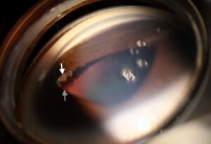
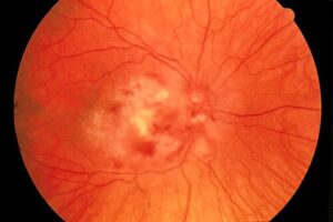
Traumatic iritis is inflammation of the iris and ciliary body that follows blunt trauma to the eye.
It is common after sports injuries, falls, and assaults, and it usually responds well to treatment.
The more important task is also to examine the eye for other injuries that can accompany it.
What Is Traumatic Iritis?
Blunt force transmits through the globe and damages the iris and ciliary body, which breaks down the blood-aqueous barrier.
Also, inflammatory cells and protein enter the anterior chamber and cause cell and flare.
The condition appears within a day or two of the injury and may also last for a week or more.
In addition, the ciliary muscle goes into spasm, causing pain, and the sphincter may go into spasm, causing miosis, or it may tear.
Clinical Features of Traumatic Iritis
- Pain, photophobia, and tearing, often a deep aching pain
- Blurred vision from ciliary spasm and cells in the anterior chamber
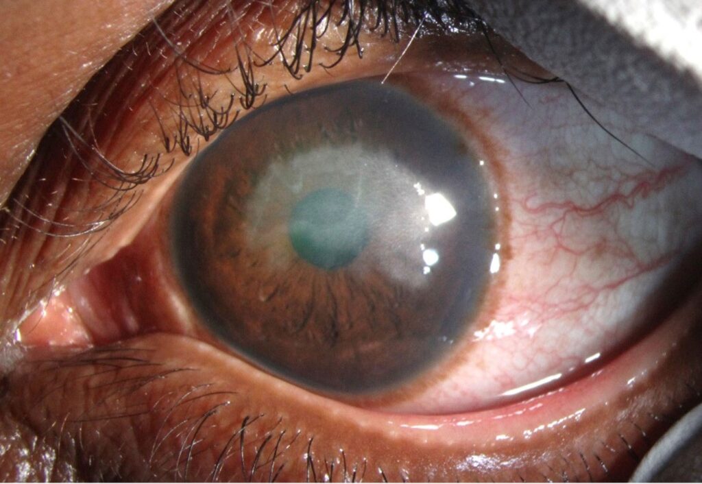
- Circumlimbal injection (ciliary flush)
- Cells and flare in the anterior chamber, and sometimes keratic precipitates
- A small, poorly reactive pupil, or a larger one with sphincter tears
- Normal or mildly reduced pressure, and sometimes a raised pressure
Also, the patient may recall a blow that happened one or two days earlier, and the symptoms may be delayed.
All-fit Slit-Lamp Adapter
Record and share exactly what you see at the slit lamp. One adapter fits any slit lamp or surgical microscope — and any smartphone.
From Choroida — the team behind this siteExamine for Other Injuries
Traumatic iritis is a marker of a significant blow, and the eye needs a thorough examination.
- Hyphema: blood in the anterior chamber (see hyphema)
- Corneal abrasion and edema (see corneal abrasion)
- Sphincter tears and iridodialysis (see traumatic mydriasis and iridodialysis)
- Lens subluxation, cataract, and a Vossius ring (see rosette-shaped cataract)
- Angle recession, with a risk of glaucoma later (see angle recession glaucoma)
- Commotio retinae, choroidal rupture, vitreous hemorrhage, and retinal tears (see commotio retinae and choroidal rupture)
- Orbital fractures and open globe injury (see ruptured globe)
Dilate the pupil once you exclude open globe injury and hyphema and examine the retina with scleral depression.
Differential Diagnosis of Traumatic Iritis
- Idiopathic or HLA-B27 anterior uveitis, which trauma can trigger or mimic (see acute anterior uveitis)
- Herpetic uveitis, which can follow minor trauma
- Sympathetic ophthalmia in penetrating injury, which is a later complication (see sympathetic ophthalmia)
- Corneal abrasion with a reactive anterior chamber
Treatment for Traumatic Iritis
- Cycloplegic drops, such as cyclopentolate 1 percent two to three times a day, to relieve ciliary spasm and prevent synechiae
- Topical corticosteroid, such as prednisolone 1 percent four times a day, tapered over one to two weeks
- Analgesia with oral agents as needed
- Rest and avoidance of strenuous activity, with eye protection
Treat the pressure if it is high, using drops, and avoid steroids if there is an active epithelial defect without antibiotic cover.
Follow-Up
Review the patient within a few days to check the response, and again at one to two weeks.
Continue to monitor the intraocular pressure, since inflammation and angle damage can cause a rise.
Perform gonioscopy a few weeks after injury, and arrange regular follow-up for years, because angle recession glaucoma can develop long after the injury.
Also, warn the patient about the symptoms of retinal detachment and instruct them to report any visual change.
Pitfalls and Special Situations
- Missing a hyphema or an open globe because the examiner hurried or used poor light
- Treating with steroids when an epithelial defect or infection is present, without antibiotic cover
- Failing to examine the posterior segment and the angle
- Not arranging follow-up for angle recession and retinal tears
- Ignoring a child who may not describe the symptoms, and consider abuse if the story does not fit
In patients with a large hyphema, the treatment changes: rest, head elevation, avoidance of aspirin, and monitoring of pressure (see hyphema).
Sports and Work Injuries
Squash balls, shuttlecocks, fists, and champagne corks are classic causes of blunt trauma, because their size allows them to strike the globe without being blocked by the orbital rim.
Protective eyewear that meets standards for the sport is the best prevention, so recommend such eyewear after any injury.
Recovery and Return to Activity
Most cases resolve within one to two weeks, with a gradual reduction in photophobia and pain.
The patient can return to normal activity once the eye is comfortable and the inflammation has resolved, but contact sports should wait until the examination is normal and the eye is cleared by the clinician.
Patients who have had significant trauma may need follow-up for years, because late complications occur.
Documentation
Record the mechanism of injury, the timing, the findings on each part of the eye, and the plan.
A clear record helps in later assessments for angle recession, cataract, and retinal problems, and it provides medico-legal protection.
For the latest studies, see the PubMed results for traumatic iritis.


Document what you see
Two smartphone imaging tools built for everyday clinic use — one for the slit lamp, one for the fundus.
From Choroida — the team behind this siteReferences
- Kuhn F, Morris R, Witherspoon CD, Mester V. The Birmingham Eye Trauma Terminology system (BETT). J Fr Ophtalmol. 2004;27:206-210.
- Salmon JF. Kanski’s Clinical Ophthalmology: A Systematic Approach. 9th ed. Elsevier; 2020.
- Yanoff M, Duker JS, eds. Ophthalmology. 5th ed. Elsevier; 2019.
- American Academy of Ophthalmology. Basic and Clinical Science Course, Section 8: External Disease and Cornea. AAO; current edition.

Writes and reviews the clinical articles on Educate. Every post is checked against peer-reviewed sources before it goes live.