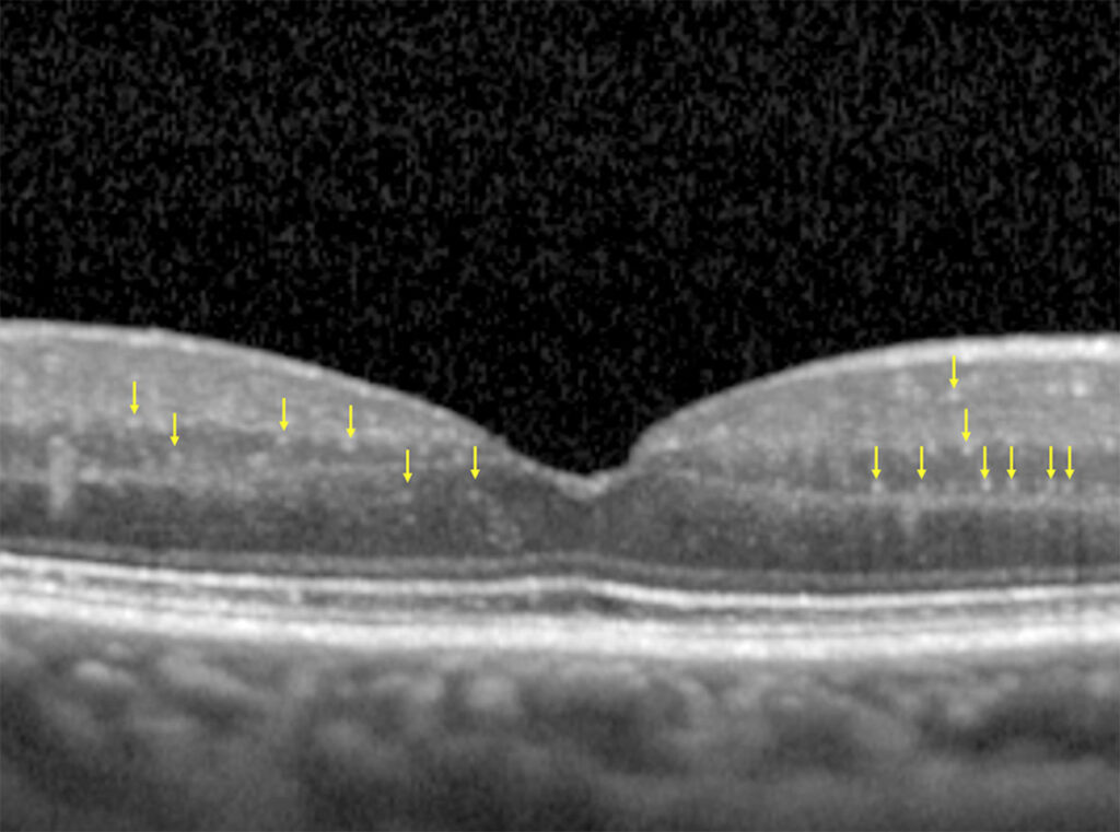
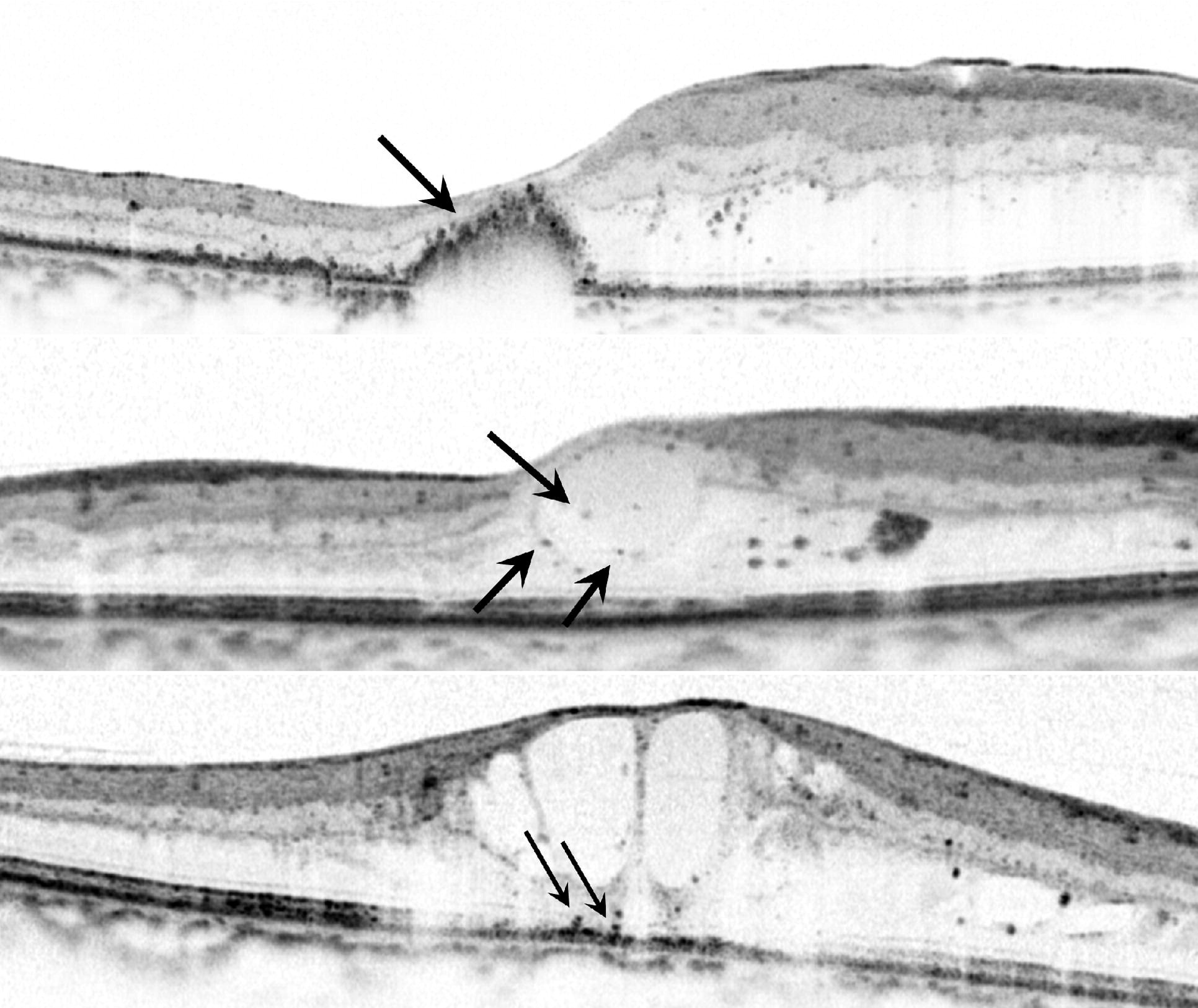
Hyperreflective foci (HRF) are small, discrete, bright dots seen within the retinal layers on OCT.
They have become one of the most discussed imaging biomarkers.
They appear in many diseases, and their number and location carry information about activity and prognosis.
Also, the meaning depends on the layer in which they are found and the disease in which they occur.
What Are Hyperreflective Foci?
HRF are punctate lesions of about 20 to 50 micrometers in size, with reflectivity similar to that of the retinal nerve fiber layer or the pigment epithelium.
They do not cast a shadow, which separates them from larger hard exudates that block deeper signals.
They can occur in any layer, but they are most often described in the inner and outer retina, and above drusen.
Because the resolution of OCT is limited, a focus may represent a cell, a cluster of cells, or a deposit.
What Do They Represent?
- Migrating retinal pigment epithelial cells, which move into the retina in age-related macular degeneration, and are linked to pigmentary changes seen on fundus examination
- Activated microglia, which are inflammatory cells that move into the outer retina, seen in diabetic retinopathy and other diseases
- Lipid-laden macrophages and extravasated lipoproteins, which are the earliest forms of hard exudate
- Degenerated photoreceptors and cell debris
Histologic correlation is limited, and several of these sources may produce the same appearance.
Fundus Explorer Pro
Photograph the retinal findings described here with the phone already in your pocket — 22 D optics and built-in illumination in one handheld unit.
From Choroida — the team behind this siteHyperreflective Foci in Diabetic Macular Edema
Bolz and colleagues described HRF in diabetic macular edema (see diabetic macular edema), and linked them to lipid extravasation, with a relationship to hard exudates (see the references).
HRF in the outer retina are also associated with a worse visual outcome and with a greater thickness of the retina.
Their number may decrease after treatment with anti-VEGF or steroids.
Reports describe a decrease with dexamethasone implant, which suggests an inflammatory component (see dexamethasone intravitreal implant).
In some studies, a high number of HRF before treatment predicted a poorer response to injections.
Hyperreflective Foci in AMD
In intermediate AMD (see age-related macular degeneration), HRF above drusen are an OCT sign that predicts progression to atrophy or neovascularization.
Christenbury and colleagues reported that progression of intermediate AMD was associated with an increase in the number of HRF (see the references).
Their presence is part of the OCT risk features, together with drusen volume and reticular pseudodrusen (see age-related macular degeneration risk factors).
In geographic atrophy, HRF at the margin of the lesion may also predict growth of atrophy.
Other Conditions
- Retinal vein occlusion: HRF accompany edema and exudates (see retinal vein occlusion workup)
- Uveitic macular edema: HRF may reflect inflammatory cells
- Macular telangiectasia and other degenerations
- Retinitis pigmentosa, with pigment migration
- Central serous chorioretinopathy, where HRF in the subretinal space may represent fibrin or inflammatory cells
How to Evaluate Them
- Look at the layer: outer retinal location and a cluster over drusen are more worrisome in AMD
- Check the number in the central macula, since some studies use counts within a defined area
- Distinguish from artifacts, such as noise, and from larger exudates
- Compare scans over time, using the same instrument
- Use them in combination with other signs, since a single marker is not enough to decide management
Developers are building automated software for counting HRF, but it is not yet standard in clinics.
Limitations of Hyperreflective Foci
- There is no standard definition, and sizes and thresholds differ
- Interobserver variability can be high
- The meaning of HRF depends on the disease and the layer, so do not apply a general rule
- Evidence on their value in treatment decisions is still limited
Practical Use
Report HRF when they are present in a scan, and note the layer and the approximate number.
Take them into account when counselling about risk, and in planning follow-up intervals, but do not treat a patient on the basis of foci alone.
Counting and Reporting
When reporting HRF, mention the layer, the number, and the distribution, for example “multiple outer retinal hyperreflective foci overlying drusen in the central macula”.
A consistent description helps the reader to compare scans and to judge the course, especially when different clinicians see the patient.
Consider that treatment may change the foci, and note whether a reduction accompanies a clinical response.
Because the origin of HRF is uncertain, avoid overstating what they mean to the patient, and describe them as a sign that the retina is under stress that warrants follow-up.
Educate trainees to look for the foci at high magnification, and to avoid mistaking speckle noise for true lesions.
For the latest studies, see the PubMed results for hyperreflective foci.


Document what you see
Two smartphone imaging tools built for everyday clinic use — one for the slit lamp, one for the fundus.
From Choroida — the team behind this siteReferences
- Bolz M, Schmidt-Erfurth U, Deak G, et al. Optical coherence tomographic hyperreflective foci: a morphologic sign of lipid extravasation in diabetic macular edema. Ophthalmology. 2009;116:914-920.
- Christenbury JG, Folgar FA, O’Connell RV, et al. Progression of intermediate age-related macular degeneration with proliferation and inner retinal migration of hyperreflective foci. Ophthalmology. 2013;120:1038-1045.
- Ryan SJ, Schachat AP, Wilkinson CP, et al, eds. Retina. 6th ed. Elsevier; 2018.
- American Academy of Ophthalmology. Basic and Clinical Science Course, Section 12: Retina and Vitreous. AAO; current edition.

Writes and reviews the clinical articles on Educate. Every post is checked against peer-reviewed sources before it goes live.