
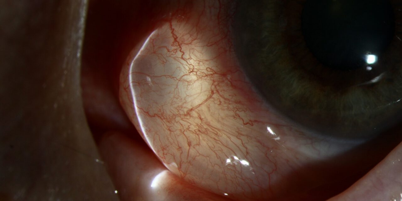
A clear, thin-walled, fluid-filled bubble sits under the conjunctiva.
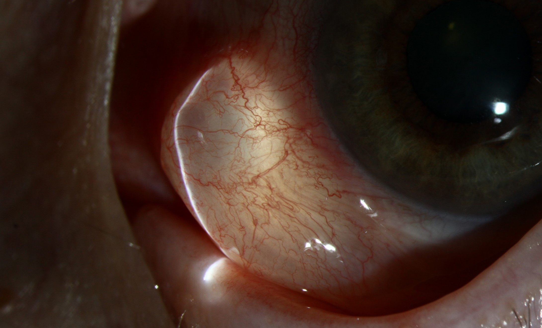
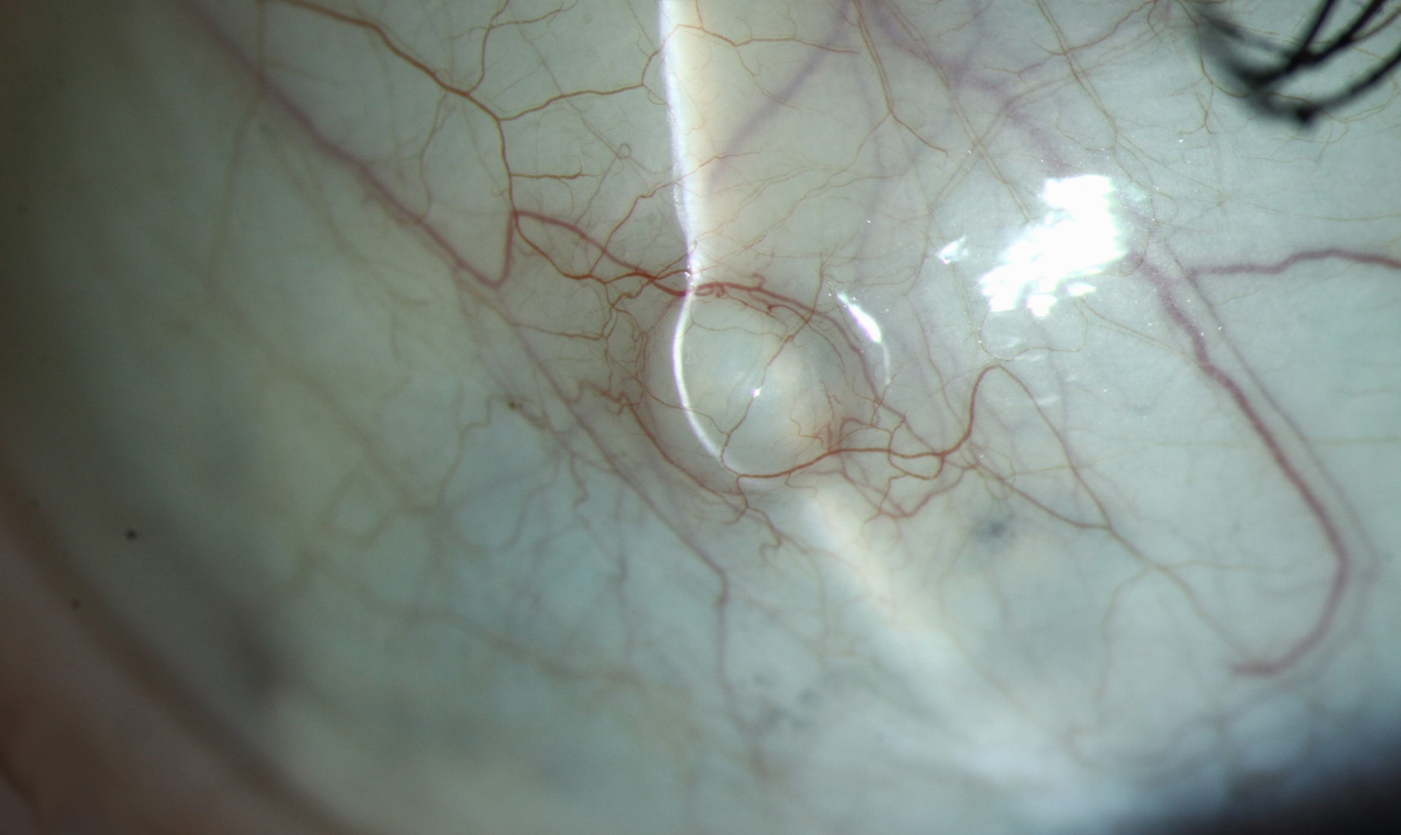
It may be a few millimeters across, sometimes larger.
It does not hurt and does not inflame, but patients notice it in the mirror or feel it when they blink.
In a majority of cases it is a conjunctival inclusion cyst.
How it forms
Conjunctival epithelium can be buried below the surface when the conjunctiva is injured, operated on, or inflamed.
The trapped cells continue to proliferate, they line a cavity, and they secrete fluid.
The cavity gradually enlarges, forming a cyst.
The commonest causes are:
- Strabismus surgery, in which the conjunctival edge may be buried
- Retinal detachment surgery with scleral buckles
- Pterygium and other ocular surface surgery
- Penetrating trauma and chronic inflammation
- Long-standing conjunctival irritation, and occasionally spontaneous cysts
Some cysts arise from glands or ducts, such as accessory lacrimal glands.
What it looks like
- A smooth, translucent, dome-shaped swelling filled with clear fluid, sometimes with a thin vascular network on the surface
- Movable with the conjunctiva, and not attached to the sclera
- Often at the site of previous surgery, or near the limbus or fornix
- Sometimes contains mucoid or slightly cloudy material, or pigment in the pigmented type
- Typically a single lesion
A thin-walled cyst may rupture and reform, leading to recurrent symptoms.
All-fit Slit-Lamp Adapter
Record and share exactly what you see at the slit lamp. One adapter fits any slit lamp or surgical microscope — and any smartphone.
From Choroida — the team behind this siteDifferential diagnosis
- Conjunctival lymphangiectasia, a chain of dilated lymphatic channels with clear fluid (see conjunctival lymphangiectasia)
- Conjunctival nevus with cystic spaces, where pigmentation is present
- Parasitic cyst, such as cysticercosis, with a visible scolex
- Conjunctival dermoid or lipodermoid, which are solid and congenital
- Conjunctival papilloma, a vascular, frond-like lesion (see conjunctival papilloma)
- Epithelial neoplasia in the rare case with an irregular, vascularized appearance
When treatment is needed
Many small cysts are asymptomatic and can be left alone.
Treatment is considered when the cyst:
- Causes discomfort, irritation, or a dry spot
- Interferes with blinking or contact lens wear
- Is cosmetically bothersome
- Is enlarging or recurrent
Treatment options
- Aspiration with a fine needle, which is simple but often leads to recurrence because the epithelial lining remains.
- Marsupialization or unroofing, followed by cautery of the base, is more reliable.
- Complete excision with care to remove the whole lining. The conjunctival defect is closed or left to heal. A cyst should be removed intact when possible, because rupture leaves cells behind.
- Cryotherapy or chemical cautery of the cyst bed may reduce recurrence.
Postoperatively, topical antibiotics and a brief course of topical steroid help to settle the eye.
Assessing the cyst in the clinic
Ask when the cyst appeared and whether there was previous surgery or injury. Examine it with a slit lamp, noting its size, translucency, vascularity, and mobility. A cyst that moves freely with the conjunctiva and is not attached to the sclera is likely to be an inclusion cyst. Transillumination shows a clear interior. Look for associated findings such as a bleb, a buried suture, or scleral buckle material, which may be the cause.
Procedure and aftercare
Under topical anesthesia, the conjunctiva over the cyst is opened and the cyst is dissected out intact if possible. The aim is to remove the lining, which prevents recurrence. If it ruptures, the surgeon removes the wall piece by piece and may touch the base with light cautery. The conjunctiva is closed with fine absorbable sutures or left to heal. Afterward, a topical antibiotic and a short course of topical steroid are used, and the patient is reviewed in a week or two.
What to tell the patient
Recurrence is possible, particularly after simple drainage, and any lesion that returns or looks different should be re-examined. Most patients are pleased with the result, and the procedure is quick and low-risk when performed carefully.
Differences from related lesions
- A lymphangiectasia is a chain of clear channels, not a single cyst, and often lies near the limbus or in the interpalpebral zone
- A pyogenic granuloma is a red, fleshy lesion after surgery
- A conjunctival nevus is pigmented and may have cystic spaces within it
- A cysticercosis cyst is a rare lesion with a visible scolex, usually in patients from endemic areas
Histology is advised when the lesion is solid, vascularized, or recurrent.
Aftercare
Advise the patient not to rub the eye, and to use the drops as directed for one to two weeks. Mild redness is normal for several days. A small number develop a recurrence in the following months, and a second procedure with wider excision is usually successful.
Prognosis
Excision gives a good result.
Recurrence may follow incomplete removal.
Any lesion that grows back, becomes vascularized, or looks atypical should be sent for histology.


Document what you see
Two smartphone imaging tools built for everyday clinic use — one for the slit lamp, one for the fundus.
From Choroida — the team behind this siteReferences
- Shields JA, Shields CL. Eyelid, Conjunctival, and Orbital Tumors: An Atlas and Textbook. 3rd ed. Philadelphia: Lippincott Williams & Wilkins; 2016.
- Krachmer JH, Mannis MJ, Holland EJ, eds. Cornea. 3rd ed. Philadelphia: Elsevier; 2011.
- Kanski JJ, Bowling B. Clinical Ophthalmology: A Systematic Approach. 7th ed. London: Elsevier; 2011.
- Yanoff M, Duker JS. Ophthalmology. 5th ed. Philadelphia: Elsevier; 2018.
Test yourself
A few questions straight from this article.
-
How does a conjunctival inclusion cyst form?
Epithelium buried below the surface after injury, surgery or inflammation keeps proliferating, lines a cavity and secretes fluid, which gradually enlarges into a cyst. -
Which operation is a common cause of conjunctival inclusion cyst because the conjunctival edge may be buried?
Strabismus surgery, in which the conjunctival edge may be buried, is listed among the commonest causes, along with retinal detachment surgery with scleral buckles. -
Which examination feature indicates a clear cystic swelling is conjunctival rather than scleral?
An inclusion cyst is movable with the conjunctiva and not attached to the sclera, which is a key feature on slit-lamp assessment. -
Why does simple needle aspiration of a conjunctival inclusion cyst often lead to recurrence?
Aspiration with a fine needle is simple but often leads to recurrence because the secreting epithelial lining is left behind. -
Which procedure is described as more reliable than aspiration for a conjunctival inclusion cyst?
Marsupialization or unroofing followed by cautery of the base is more reliable than aspiration, and complete excision of the whole lining is also used. -
Which topical regimen is used after excision of a conjunctival inclusion cyst?
Postoperatively topical antibiotics and a brief course of topical steroid are used to help settle the eye. -
How does conjunctival lymphangiectasia differ in form from a conjunctival inclusion cyst?
Lymphangiectasia appears as a chain of dilated lymphatic channels containing clear fluid, whereas an inclusion cyst is typically a single lesion. -
Which feature identifies a parasitic conjunctival cyst such as cysticercosis?
A parasitic cyst such as cysticercosis is recognised by a visible scolex, usually in patients from endemic areas. -
Which problem is an accepted indication to treat a conjunctival inclusion cyst?
Treatment is considered when the cyst causes discomfort, irritation or a dry spot, interferes with blinking or contact lens wear, is cosmetically bothersome, or is enlarging or recurrent. -
When should a presumed conjunctival inclusion cyst be sent for histology?
Histology is advised when the lesion is solid, vascularized or recurrent, and any lesion that grows back or looks atypical should be sent for examination.
