
Tortuous retinal vessels, arteries or veins that follow an abnormally winding, curved course rather than their normal, relatively straight radial path from the optic disc, are a fundus finding with a genuinely broad differential diagnosis, and working through that differential systematically, by asking which vessels are involved, whether the change is generalized or localized, and what other findings accompany it, is what turns this single sign into a useful diagnostic starting point.
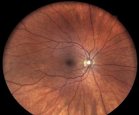
What Normal Retinal Vessels Look Like
Retinal blood vessels normally course from the optic disc toward the periphery in a relatively direct, gently curving path, and while a degree of normal variation exists between individuals, a vessel pattern that is conspicuously more winding, curved, or corkscrew-like than expected is what defines tortuosity as a clinical finding worth further evaluation.
Venous Tortuosity
Systemic Causes
- Hypertensive retinopathy, in which chronic elevated blood pressure can produce venous tortuosity alongside its other characteristic findings, including arteriolar narrowing and, in more severe cases, hemorrhages and cotton wool spots
- Hyperviscosity syndromes, including polycythemia and paraproteinemias such as Waldenström macroglobulinemia, classically produce marked venous dilation and tortuosity, sometimes described as having a “sausage-link” appearance, reflecting the underlying increased blood viscosity
- Diabetic retinopathy, where venous beading and tortuosity are recognized features, particularly in more advanced, non-proliferative disease, and factor into standard diabetic retinopathy severity grading
Localized Causes
- Venous tortuosity localized to a specific retinal quadrant can indicate venous outflow obstruction in that distribution, including branch retinal vein occlusion, where tortuosity develops downstream of the occlusion as the vessel adapts to altered flow dynamics
Fundus Explorer Pro
Photograph the retinal findings described here with the phone already in your pocket — 22 D optics and built-in illumination in one handheld unit.
From Choroida — the team behind this siteArterial Tortuosity
Arterial tortuosity is less common than venous tortuosity but carries its own specific associations.
- Familial retinal arteriolar tortuosity, a rare, generally benign, autosomal dominant condition in which arterial tortuosity is an isolated finding, notable for a predisposition to recurrent retinal hemorrhage, sometimes triggered by minor trauma or Valsalva-type maneuvers, without other significant retinal pathology
- Some congenital and connective tissue disorders affecting vascular wall integrity can produce arterial tortuosity as one of several vascular manifestations
Congenital Vascular Tortuosity in Retinopathy of Prematurity and Related Conditions
Increased vascular tortuosity, particularly affecting the posterior pole vessels, is a specifically important finding in retinopathy of prematurity, where “plus disease,” defined in part by a defined degree of posterior vascular tortuosity and dilation, indicates more severe, actively progressing disease requiring urgent treatment consideration.
Tortuous, abnormally developed retinal vessels are also a feature of familial exudative vitreoretinopathy, where the vascular tortuosity accompanies other signs of abnormal peripheral retinal vascular development (see familial exudative vitreoretinopathy).
Clinical Evaluation
- Careful characterization of whether tortuosity affects arteries, veins, or both, and whether it is generalized throughout the fundus or localized to a specific area, since this distinction substantially narrows the differential
- Blood pressure measurement and assessment for other signs of hypertensive retinopathy when tortuosity is generalized and venous-predominant
- Complete blood count and, when clinically indicated, serum protein studies to evaluate for an underlying hyperviscosity syndrome when tortuosity is marked and accompanied by other suggestive fundus findings
- A family history and examination for other affected relatives when an isolated, otherwise unexplained tortuosity pattern suggests a hereditary vascular condition
- In a premature infant, careful, systematic assessment of posterior vascular tortuosity is a core, standardized component of retinopathy of prematurity screening and staging
Management
Management is directed entirely at the underlying cause once identified, since tortuous vessels themselves are a sign rather than a disease process requiring direct treatment.
- Hypertensive and hyperviscosity-related tortuosity improves with control of the underlying systemic condition
- Familial retinal arteriolar tortuosity, being generally benign, is managed with patient education about the small risk of recurrent hemorrhage and reassurance, without disease-specific treatment
- Tortuosity as a marker of disease severity in retinopathy of prematurity directly informs treatment timing decisions for that condition, given its role in defining sight-threatening “plus disease”
Prognosis
The prognosis associated with tortuous retinal vessels depends entirely on the underlying cause, ranging from an essentially benign, isolated familial finding requiring only reassurance to a marker of severe, treatment-urgent disease in the case of plus disease in retinopathy of prematurity.
This wide range of possible significance is exactly why tortuosity, recognized during a routine fundus examination, deserves systematic evaluation of its pattern and associated findings rather than being noted in isolation without further consideration of what it might represent.
Related Reading


Document what you see
Two smartphone imaging tools built for everyday clinic use — one for the slit lamp, one for the fundus.
From Choroida — the team behind this siteReferences
- Sun C, Wang JJ, Mackey DA, Wong TY. Retinal vascular caliber: systemic, environmental, and genetic associations. Surv Ophthalmol. 2009;54:74-95.
- International Committee for the Classification of Retinopathy of Prematurity. The International Classification of Retinopathy of Prematurity revisited. Arch Ophthalmol. 2005;123:991-999.
- Sears JE, Pietz J, Sonnie C, Dolcini D, Hoppe G. A change in diagnostic approach to retinopathy of prematurity based on plus disease. J AAPOS. 2009;13:344-348.
- Wallace RT, Crouch ER. Familial retinal arteriolar tortuosity. J Pediatr Ophthalmol Strabismus. 1992;29:145-150.
