
After LASIK, the corneal flap is replaced and heals to the underlying stroma.
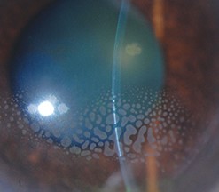
In some patients, a few epithelial cells slip beneath the flap edge and start to grow in the interface.
This is epithelial ingrowth, and it is one of the more important late complications after flap-based refractive surgery, because it can be self-limited and harmless or, in the worst case, destroy the flap.
How it happens
The edge of the flap does not seal like the original tissue.
Epithelium can enter the interface through a gap at the edge, through a buttonhole in the flap, or on the underside of a lifted flap during an enhancement.
Once the cells are inside, they proliferate in nests or sheets, and if the flap is not tightly adherent, the growth progresses.
Epithelial cells also release enzymes that can digest the stromal collagen around them, and this explains the risk of flap melt in more aggressive cases.
Risk factors
- Enhancement procedures, in which the flap is lifted a second time, which is the most important risk factor
- Flap buttonholes, tears, or irregular edges
- Corneal trauma after surgery
- Epithelial defects at the time of surgery, or a poorly adherent flap
- Hyperopic LASIK, where a larger flap or a wider ablation is used
- Older age, diabetes, and recurrent erosion
Clinical picture
- Many cases are found incidentally at a routine check, as small, grayish-white, translucent islands at the peripheral flap edge, with no symptoms
- Progressive cases produce blur, glare, irregular astigmatism, and sometimes pain or foreign-body sensation
- Under the slit lamp, there are pearls or sheets of translucent cells with a sharp, rounded edge, which are best seen with retroillumination, and there may be a whitish line at the leading edge
- In advanced cases, the overlying flap thins and melts, with stromal haze and scarring
Differential diagnosis
- Diffuse lamellar keratitis, a sterile inflammation that develops in the first days, with granular, hazy deposits (see diffuse lamellar keratitis)
- Infectious keratitis, which has an infiltrate, pain, and redness
- Interface debris or fibers, which are non-progressive
- Striae and flap folds, seen as linear lines
- Pressure-induced interface fluid in patients with elevated intraocular pressure
Management
- Observation is appropriate for small, peripheral, non-progressive ingrowth that does not threaten the visual axis or flap integrity, with regular follow-up and documentation by photographs.
- Surgical removal is needed for progressive ingrowth, visual disturbance, flap melt, or involvement near the visual axis. The surgeon lifts the flap, scrapes the epithelium from both the stromal bed and the underside of the flap, and replaces the flap carefully. Sutures, fibrin glue, or a bandage lens may be used to prevent recurrence.
- Nd:YAG laser has been used to ablate small nests.
- Recurrent cases may need repeat treatment with additional measures, such as suturing the flap edge.
Prevention
Careful flap creation and repositioning, minimal manipulation, avoiding epithelial defects, and caution at the time of enhancement are important.
Surgeons often prefer surface ablation for enhancement when the flap is old, to avoid lifting it.
How to find it
Examine the flap edges with a slit lamp, using direct illumination and retroillumination. Epithelial cells look like translucent, pearl-like clusters or sheets, with sharp margins and sometimes a fine whitish line at the border. Compare with previous visits, because the key question is whether the lesion has grown. Corneal topography may show localized steepening or irregular astigmatism over an area of ingrowth, and anterior segment OCT shows the cells as a hyperreflective layer in the interface.
Grading for decisions
Surgeons often grade ingrowth by the distance it has advanced from the flap edge and by its effect on the flap. Small areas within 1 to 2 mm of the edge that are stable can be watched. Areas that grow, that reach the visual axis, that cause flap thinning, or that cause symptoms need to be removed. Recurrent ingrowth after removal suggests a persistent entry point, and the surgeon may suture the flap or use glue.
Counselling
Explain to the patient that ingrowth is rare, that it can be treated, and that early detection helps to prevent damage. Patients who have had enhancement procedures or flap complications should be told to attend regular follow-up and to report blur, glare, or discomfort.
Documenting and sharing the finding
Record the location, size, and distance from the visual axis, and take an image. If you are not the operating surgeon, send a note to the surgeon, since the history of the flap and the enhancements is important in deciding on management.
Prognosis
Most patients have a good outcome with timely treatment.
Neglected, advanced cases can leave scars and irregular astigmatism.


Document what you see
Two smartphone imaging tools built for everyday clinic use — one for the slit lamp, one for the fundus.
From Choroida — the team behind this siteReferences
- Wang MY, Maloney RK. Epithelial ingrowth after laser in situ keratomileusis. Am J Ophthalmol. 2000;129:746-751.
- Ting DSJ, Srinivasan S, Danjoux JP. Epithelial ingrowth following laser in situ keratomileusis (LASIK): prevalence, risk factors, management and visual outcomes. BMJ Open Ophthalmol. 2018;3:e000133.
- Ambrosio R Jr, Wilson S. Complications of laser in situ keratomileusis: etiology, prevention, and treatment. J Refract Surg. 2001;17:350-379.
- Krachmer JH, Mannis MJ, Holland EJ, eds. Cornea. 3rd ed. Philadelphia: Elsevier; 2011.
