
IOL glistenings are tiny, fluid-filled microvacuoles that form inside the optic of some hydrophobic acrylic intraocular lenses, and they shine like glittering droplets under the slit lamp.
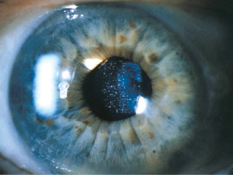
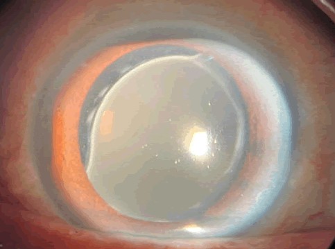
They are common in lenses of this material, they increase with time, and in most patients they have no effect on vision.
The clinical question is when they matter, and how to separate them from opacities that do.
What Are IOL Glistenings?
Hydrophobic acrylic lenses absorb a small amount of water when they sit in the aqueous humor.
When the temperature of the lens changes, water that has entered the polymer can separate into microscopic pockets, and these pockets scatter light.
The vacuoles are typically 1 to 20 micrometers in diameter, and they may be seen in the central optic or throughout it.
They tend to appear months to years after surgery, and their density increases with time and with exposure to temperature changes.
The material, the manufacturing process, and the packaging influence how many form, and newer lens materials have been designed to reduce them.
Clinical Features
Most patients are asymptomatic, and the finding is discovered at the slit lamp.
In direct illumination, the vacuoles appear as small, bright, refractile points in the lens optic, and with retroillumination they look dark.
Dense glistenings may be associated with a mild decrease in contrast sensitivity and a feeling of glare or haze, particularly at night.
The visual acuity measured on a chart is often normal, which is why the complaint may seem out of proportion to the examination.
The vacuoles are in the lens itself and do not move with the position of the eye, and they cannot be removed with laser capsulotomy.
All-fit Slit-Lamp Adapter
Record and share exactly what you see at the slit lamp. One adapter fits any slit lamp or surgical microscope — and any smartphone.
From Choroida — the team behind this siteDifferential Diagnosis
- Calcification of hydrophilic lenses: granular white deposits on or within the lens, with a different material and time course
- Posterior capsule opacification: haze behind the lens that responds to laser capsulotomy (see YAG laser capsulotomy)
- Pigment dispersion on the lens surface: fine brown dots on the anterior surface that can be wiped off
- Inflammatory precipitates: cells and fibrin on the lens surface in uveitis
- Surface light scattering: fine, whitish changes on the optic surface seen with certain lens materials
A careful slit-lamp examination that focuses first on the anterior surface, then the optic, and then the posterior capsule usually separates these.
Evaluation
Document the density with a grading scale and a photograph, and measure the best-corrected acuity and the contrast sensitivity.
Ask about glare, halos, and night vision, and try to relate the symptoms to the density of the vacuoles.
Look at the other eye, because the problem is often bilateral and similar.
Rule out other causes of reduced contrast, including macular disease, posterior capsule opacification, and dry eye, before attributing symptoms to glistenings.
Management
Observation is appropriate in the great majority of eyes.
Patients should be reassured that the vacuoles are within the lens material, that they are not a sign of infection or inflammation, and that they usually remain stable.
In the very small number of patients with symptoms that are clearly related to dense glistenings, a lens exchange can be discussed, but the risks of surgery must be weighed against the benefit.
Laser techniques to reduce the vacuoles have been described in laboratory and early clinical studies, but they are not standard care.
Any lens exchange should follow the usual principles for explantation, and the surgeon should consider the status of the capsule and the zonules (see capsular tension ring indications).
Record the lens model and the date of surgery in the chart, because this helps in later decisions.
Counselling and Documentation
Patients who read about glistenings on the internet may worry that the lens is failing.
A clear explanation that the vacuoles are tiny, that they sit within the optic material, and that the retina and optic nerve are unaffected reassures most patients.
Make a note of the lens model in the record, together with the grade of the glistenings and the date of the examination.
This note is valuable if the patient later develops decreased contrast or night vision problems, because it shows the finding was present and stable.
Dense glistenings tend to be bilateral and symmetrical, and a unilateral dense change should prompt a search for another cause.
Patients with persistent complaints and a normal examination can be tested for contrast sensitivity, which may confirm the deficit and help decide on management.
Newer hydrophobic acrylic materials have been engineered to produce fewer vacuoles, so the severity depends partly on the lens platform that was used.
When a patient is planning a second eye operation after developing symptoms in the first, discuss the lens choice with this history in mind.


Document what you see
Two smartphone imaging tools built for everyday clinic use — one for the slit lamp, one for the fundus.
From Choroida — the team behind this siteReferences
- Werner L. Glistenings and surface light scattering in intraocular lenses. J Cataract Refract Surg. 2010;36:1398-1420.
- Miyata A, Yaguchi S. Equilibrium water content and glistenings in acrylic intraocular lenses. J Cataract Refract Surg. 2004;30:1768-1772.
- Salmon JF. Kanski’s Clinical Ophthalmology: A Systematic Approach. 9th ed. Elsevier; 2020.
- American Academy of Ophthalmology. Basic and Clinical Science Course, Section 11: Lens and Cataract. AAO; current edition.

Writes and reviews the clinical articles on Educate. Every post is checked against peer-reviewed sources before it goes live.


