
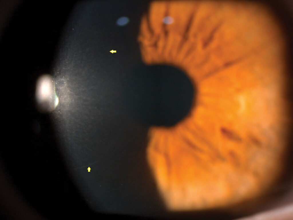
Corneal nerve imaging with in vivo confocal microscopy allows direct, non-invasive visualization of the sub-basal nerve plexus, the dense network of corneal nerves lying just beneath the epithelium, and it has turned corneal nerve assessment from something inferred indirectly through sensation testing into something that can be measured and quantified in living tissue, in real time, in the clinic.
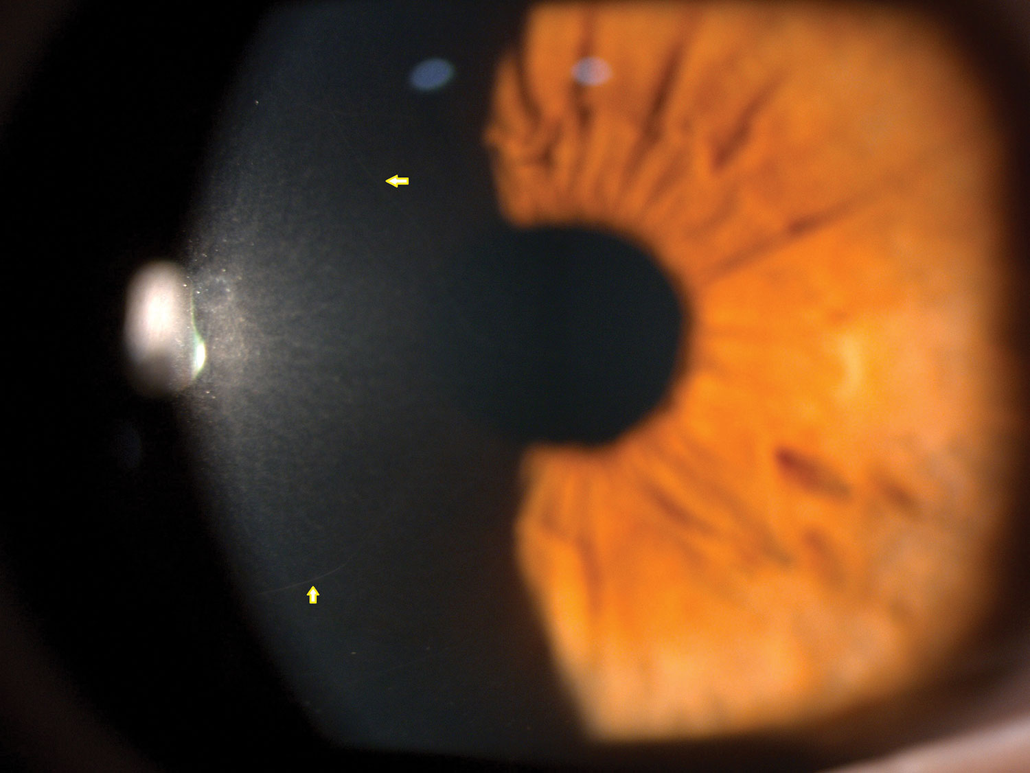
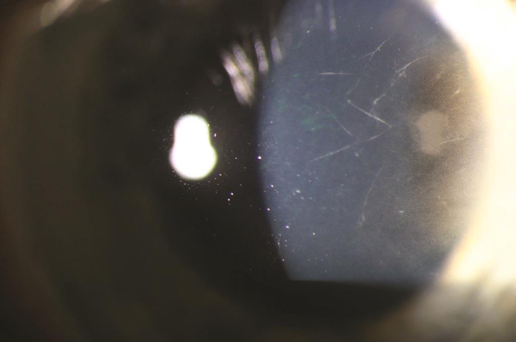
What In Vivo Confocal Microscopy Shows
In vivo confocal microscopy uses a laser scanning system to image the cornea at the cellular level, layer by layer, without the need for a biopsy.
At the level of the sub-basal nerve plexus, it visualizes individual nerve fibers as bright, linear structures running roughly parallel to Bowman layer, and specific parameters, including nerve fiber density, nerve branch density, and nerve fiber length, can be measured and tracked over time as objective, quantitative markers of corneal nerve health.
Why Corneal Nerves Are a Useful Window
The cornea is one of the most densely innervated tissues in the body, and its nerves are accessible to direct, non-invasive imaging in a way that peripheral nerves elsewhere in the body are not, which has made corneal nerve imaging a research tool for studying systemic small-fiber neuropathy, not only for corneal disease specifically.
Reduced corneal nerve density and altered nerve morphology have been documented in diabetes, sometimes preceding clinically detectable peripheral neuropathy elsewhere in the body, raising interest in corneal confocal microscopy as an early biomarker for diabetic neuropathy more broadly, beyond its direct relevance to corneal health.
All-fit Slit-Lamp Adapter
Record and share exactly what you see at the slit lamp. One adapter fits any slit lamp or surgical microscope — and any smartphone.
From Choroida — the team behind this siteClinical Applications
Diabetic Corneal Neuropathy
Diabetes damages small corneal nerve fibers, often before other clinically apparent complications, and confocal microscopy can detect reduced nerve density and altered nerve morphology in diabetic patients, correlating to some degree with the severity of diabetic peripheral neuropathy elsewhere in the body and with duration and control of diabetes.
This underlies the increased risk of neurotrophic keratitis and delayed epithelial healing seen in diabetic patients, particularly after ocular surgery (see neurotrophic keratitis).
Dry Eye Disease
Corneal nerve changes, including altered nerve density and morphology, have been documented in various forms of dry eye disease, and some patients with significant dry eye symptoms but relatively mild signs on conventional examination show measurable nerve abnormalities on confocal imaging, supporting a neuropathic component to their symptoms.
Post-Refractive Surgery and Post-Herpetic Evaluation
Confocal microscopy can track corneal nerve regeneration after procedures that transect corneal nerves, such as LASIK, and after herpetic keratitis, providing objective evidence of nerve recovery, or its absence, relevant to assessing ongoing neurotrophic risk.
Monitoring Treatment Response
For conditions such as neurotrophic keratitis being treated with nerve growth factor or other regenerative therapies, serial confocal imaging can provide objective evidence of nerve regeneration alongside clinical improvement in sensation and epithelial healing.
Practical Considerations
In vivo confocal microscopy requires specialized equipment and training to acquire and interpret images reliably, and it is not yet in routine widespread clinical use outside of specialized corneal and dry eye centers, though its research applications have expanded substantially and clinical availability continues to grow.
Image acquisition can be affected by patient cooperation and corneal surface irregularity, and interpretation benefits from experience given normal variation in nerve patterns between individuals and across different corneal regions.
Future Directions
Standardization of imaging protocols and normative nerve density data across populations remains an active area of work, which would support broader clinical adoption of corneal nerve density as a routine biomarker, both for primary corneal disease and as a window into systemic small-fiber neuropathy more generally.
Prognosis and Clinical Value
Corneal nerve imaging does not treat disease itself, but it provides objective, quantitative information that can support earlier diagnosis of corneal neuropathy, guide treatment decisions in neurotrophic and dry eye disease, and track response to nerve-directed therapies over time in a way that symptom-based assessment alone cannot.


Document what you see
Two smartphone imaging tools built for everyday clinic use — one for the slit lamp, one for the fundus.
From Choroida — the team behind this siteReferences
- Patel DV, McGhee CN. Contemporary in vivo confocal microscopy of the living human cornea using white light and laser scanning techniques: a major review. Clin Exp Ophthalmol. 2007;35:71-88.
- Tavakoli M, Petropoulos IN, Malik RA. Corneal confocal microscopy to assess diabetic neuropathy: an eye on the foot. J Diabetes Sci Technol. 2013;7:1179-1189.
- Labbe A, Liang Q, Wang Z, et al. Corneal nerve structure and function in patients with non-Sjogren dry eye: clinical correlations. Invest Ophthalmol Vis Sci. 2013;54:5144-5150.
- Cruzat A, Qazi Y, Hamrah P. In vivo confocal microscopy of corneal nerves in health and disease. Ocul Surf. 2017;15:15-47.

Writes and reviews the clinical articles on Educate. Every post is checked against peer-reviewed sources before it goes live.


