
Lisch epithelial corneal dystrophy (LECD) is an X-linked dominant dystrophy of the corneal epithelium, characterized by clusters of tiny intraepithelial cysts arranged in bands and whorls.
It was described by Lisch and colleagues in 1992 in a single family, and it is now recognized as a distinct entity in the IC3D classification of corneal dystrophies.
Most patients have good vision, and the diagnosis is often made on a routine slit-lamp examination.
What Lisch Epithelial Corneal Dystrophy Looks Like
The opacities are small, feathery, gray clusters in the epithelium, and they tend to form band-shaped or whorled patterns that sweep across the cornea.
Between the bands the cornea is clear, and the stroma and endothelium are normal.
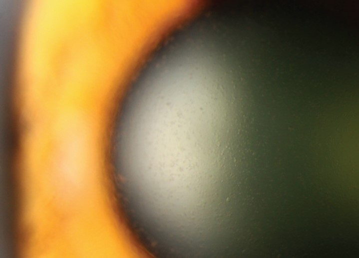
Retroillumination is the most useful technique, because it shows the cysts as clear, round vacuoles against the red reflex, and it separates them from deposits and scars.
In direct illumination the same areas look like a faint, patchy haze.
The pattern can be bilateral and asymmetric, and the distribution varies a great deal from eye to eye.
Genetics
The condition maps to the Xp22.3 region, and it follows an X-linked dominant pattern with variable expression.
The causative gene has not been firmly established, so molecular confirmation is not routinely available.
Males tend to be affected more uniformly, and females often show band or whorl patterns that are thought to reflect random X inactivation.
A family history on the maternal side, with an affected son and mother, is a useful clue when it is present.
All-fit Slit-Lamp Adapter
Record and share exactly what you see at the slit lamp. One adapter fits any slit lamp or surgical microscope — and any smartphone.
From Choroida — the team behind this siteSymptoms and Course
Many patients have no symptoms, and the finding is discovered by chance.
Some report mild blur, glare, or fluctuating vision, and these complaints are more likely when the cysts involve the visual axis or cause irregular astigmatism.
Recurrent erosions are uncommon, but they have been described, and they should be treated like any other erosion (see recurrent corneal erosion).
The disease is usually stable, and progression is slow if it happens at all.
Differential Diagnosis
- Meesmann dystrophy: tiny, uniform, diffuse epithelial cysts without a band or whorl arrangement (see Meesmann corneal dystrophy)
- Epithelial basement membrane dystrophy: maps, dots, and fingerprint lines, with a fluctuating course (see map-dot-fingerprint dystrophy)
- Vortex keratopathy: a whorl of subepithelial deposits from amiodarone, chloroquine, or Fabry disease (see cornea verticillata)
- Contact lens epithelial changes: microcysts that appear after lens wear and recede after discontinuation
The whorled pattern invites confusion with vortex keratopathy, but in LECD the opacities are intraepithelial cysts and not pigmented or golden deposits.
A medication list and a family history resolve most doubtful cases.
Management
Observation is appropriate when vision is good and the patient is comfortable.
Lubricants help with fluctuating vision, and a bandage lens or hypertonic ointment can be used if erosions occur.
When the cysts cause visual symptoms, superficial keratectomy or phototherapeutic keratectomy can clear the epithelium, although recurrence is possible and the effect may not last.
Patients should be told that the condition is inherited, that relatives may be affected, and that it does not threaten the eye in the way that a stromal or endothelial dystrophy can.
Documentation with retroillumination photographs gives a baseline for any later comparison.
Imaging and Documentation
Anterior segment OCT shows the cysts as small, round, hyporeflective spaces within the epithelial layer, and it confirms that the stroma beneath is normal.
In vivo confocal microscopy can show the intraepithelial microcysts as dark, rounded spaces with a thin bright border, and the surrounding basal epithelial cells may look slightly irregular.
These techniques are not needed in a typical case, but they are useful when the clinical picture is atypical or when the clinician wants an objective baseline.
Corneal topography is worth obtaining if the patient has visual complaints, because irregular astigmatism from epithelial thickening can be quantified.
A Practical Note on Family Screening
Because the inheritance is X-linked dominant, an affected father cannot pass the condition to a son, whereas an affected mother can pass it to children of either sex.
Examining a parent or a sibling who is available takes a few minutes and often confirms the diagnosis, since relatives may be unaware that they have the same opacities.
Female carriers may show only a subtle whorl, so the examination should include retroillumination and a dilated pupil when the pattern is faint.
A short record of the pattern, the laterality, and the family history in the chart saves time at later visits, particularly when a different clinician sees the patient and wonders whether the opacities are new.


Document what you see
Two smartphone imaging tools built for everyday clinic use — one for the slit lamp, one for the fundus.
From Choroida — the team behind this siteReferences
- Lisch W, Steuhl KP, Lisch C, et al. A new, band-shaped and whorled microcystic dystrophy of the corneal epithelium. Am J Ophthalmol. 1992;114:35-44.
- Weiss JS, Møller HU, Aldave AJ, et al. IC3D classification of corneal dystrophies, edition 2. Cornea. 2015;34:117-159.
- Mannis MJ, Holland EJ, eds. Cornea. 4th ed. Elsevier; 2017.
- American Academy of Ophthalmology. Basic and Clinical Science Course, Section 8: External Disease and Cornea. AAO; current edition.

Writes and reviews the clinical articles on Educate. Every post is checked against peer-reviewed sources before it goes live.


