
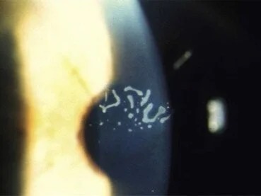
Map-dot-fingerprint dystrophy, formally known as epithelial basement membrane dystrophy, is the most common of the anterior corneal dystrophies.
Its practical significance lies far less in any direct effect on vision than in its role as the single most common identifiable underlying cause of recurrent corneal erosion syndrome.
Many patients live with the condition entirely unaware of it until a first episode of erosion brings them in for evaluation.
Pathogenesis
The disease reflects abnormal production of basement membrane material by the corneal epithelium, resulting in redundant, poorly organized basement membrane that extends up into the epithelial layer itself rather than staying in its normal, thin position beneath it.
This abnormal, thickened, and irregular basement membrane provides weaker, less reliable adhesion between the epithelium and the underlying Bowman’s layer, which is the direct structural link to why these patients are so prone to recurrent epithelial erosion.
The connection point itself is structurally unsound, even in areas that have never suffered a prior traumatic injury.
Most cases are sporadic and considered a degenerative rather than strictly inherited condition, though some families show an autosomal dominant pattern, and the underlying basement membrane abnormality tends to become more apparent and more widespread with advancing age.
Clinical Patterns
The condition takes its descriptive name from the three patterns the abnormal basement membrane material can take on exam, though a single patient can show more than one pattern simultaneously or may show a subtler variant that is easy to overlook without specifically looking for it:
- Map pattern — irregular, geographic-appearing gray lines outlining patches of abnormal epithelium
- Dot pattern — small, round or oval, gray-white intraepithelial microcysts, sometimes called Cogan microcysts
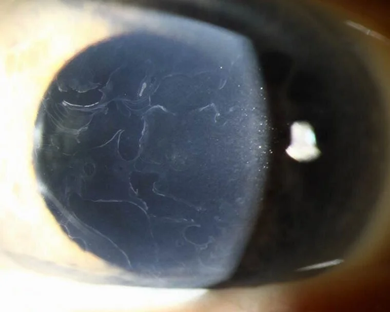
- Fingerprint pattern — fine, curvilinear parallel lines resembling a fingerprint, often best seen with retroillumination or broad tangential illumination at the slit lamp
Fundus Explorer Pro
Photograph the retinal findings described here with the phone already in your pocket — 22 D optics and built-in illumination in one handheld unit.
From Choroida — the team behind this siteClinical Presentation
A substantial proportion of patients are entirely asymptomatic and the dystrophy is discovered incidentally during a routine exam performed for another reason, because the changes themselves do not typically cause significant symptoms directly.
When symptomatic, patients present either with recurrent corneal erosion episodes — sudden, sharp pain typically on waking, discussed in more detail in this site’s dedicated article on that syndrome — or, less commonly, with mild, fluctuating blur from irregular astigmatism when the changes happen to involve the central visual axis densely enough to distort the corneal surface.
Because symptoms are so often absent or mild between erosion episodes, patients frequently do not connect their recurrent morning eye pain to an underlying, persistent corneal finding, and are sometimes surprised to learn during a quiet-eye examination that a specific, named condition explains what had previously seemed like a series of unrelated, unexplained episodes.
Exam Findings
- One or more of the map, dot, or fingerprint patterns described above, most easily appreciated with broad, tangential slit-lamp illumination or retroillumination against the red reflex
- Often bilateral, though the density and pattern can be asymmetric between the two eyes
- Subtle changes can be genuinely easy to miss on routine direct illumination alone, which is why specifically using retroillumination is worthwhile whenever recurrent erosion is being investigated
- Fluorescein staining or an active epithelial defect only during an acute erosion episode, otherwise the surface between episodes can appear relatively unremarkable on casual inspection
Differential Diagnosis
- Recurrent corneal erosion from a prior traumatic injury without underlying dystrophy — a purely traumatic history without the bilateral map/dot/fingerprint findings characteristic of the dystrophy
- Meesmann corneal dystrophy — also produces intraepithelial microcysts, but characteristically presents earlier in life (childhood) and shows a different, more uniform distribution across the entire cornea rather than the patchy, variable pattern of map-dot-fingerprint dystrophy
- Other anterior corneal dystrophies (Reis-Bücklers, and others) — generally rarer, with their own distinct patterns on careful slit-lamp exam
Diagnostic Evaluation
The diagnosis is made clinically, based on the characteristic map, dot, or fingerprint pattern identified with careful slit-lamp exam, ideally using broad tangential illumination or retroillumination to bring out changes that are easily missed with routine direct illumination alone.
Corneal topography can be useful when irregular astigmatism from central involvement is contributing to visual symptoms, helping to characterize and quantify the degree of surface irregularity present.
No further testing is typically needed once the characteristic pattern is identified and the history of recurrent, self-limited erosion episodes, when present, fits the expected clinical picture, though a persistently atypical or non-healing epithelial defect should prompt broader consideration of other causes rather than being assumed to be a straightforward dystrophy-related erosion.
Management
Asymptomatic patients require no treatment beyond being informed of the finding, because it does not by itself threaten vision or require intervention.
Patients with recurrent erosion related to the dystrophy are managed following the same principles described in this site’s dedicated article on recurrent corneal erosion syndrome: aggressive lubrication as first-line prevention, with hypertonic saline, anterior stromal micropuncture, diamond burr epithelial debridement, or phototherapeutic keratectomy reserved for cases refractory to lubrication alone.
Because the underlying basement membrane abnormality is diffuse rather than confined to a single prior injury site, patients with map-dot-fingerprint dystrophy who undergo any of these more definitive interventions should be counseled that the fellow eye, or other areas of the same eye, may still develop erosion in the future.
This is because the procedure addresses the treated area specifically rather than curing the underlying, more widespread dystrophic tendency.


Document what you see
Two smartphone imaging tools built for everyday clinic use — one for the slit lamp, one for the fundus.
From Choroida — the team behind this siteReferences
- Laibson PR. Recurrent corneal erosions and epithelial basement membrane dystrophy. Eye & Contact Lens.
- Werblin TP, Hirst LW, Stark WJ, Maumenee IH. Prevalence of map-dot-fingerprint changes in the cornea. British Journal of Ophthalmology.
- American Academy of Ophthalmology. Basic and Clinical Science Course, Section 8: External Disease and Cornea.
Test yourself
A few questions straight from this article.
-
Map-dot-fingerprint dystrophy is formally known by which other name?
Map-dot-fingerprint dystrophy is the formal entity epithelial basement membrane dystrophy, and it is the most common of the anterior corneal dystrophies. -
Map-dot-fingerprint dystrophy is the most commonly identified underlying cause of which condition?
The practical importance of the dystrophy lies in its role as the single most common identifiable underlying cause of recurrent corneal erosion syndrome, rather than in any direct effect on vision. -
In map-dot-fingerprint dystrophy, abnormal basement membrane weakens epithelial adhesion to which structure?
The thickened, irregular basement membrane provides weaker adhesion between the epithelium and the underlying Bowman's layer, which explains the tendency to recurrent epithelial erosion. -
What inheritance pattern applies to most cases of map-dot-fingerprint dystrophy?
Most cases are sporadic and regarded as degenerative, although some families do show an autosomal dominant pattern and the changes become more widespread with age. -
The intraepithelial microcysts seen in map-dot-fingerprint dystrophy are also called:
The dot pattern consists of small round or oval gray-white intraepithelial microcysts, sometimes referred to as Cogan microcysts. -
Which slit-lamp technique best reveals the fine fingerprint lines of map-dot-fingerprint dystrophy?
The curvilinear fingerprint lines are often invisible on routine direct illumination and are best appreciated with retroillumination against the red reflex or broad tangential illumination. -
Which dystrophy also causes intraepithelial microcysts but appears in childhood with uniform corneal distribution?
Meesmann dystrophy also produces intraepithelial microcysts, but it presents earlier in childhood and shows a more uniform distribution across the whole cornea instead of the patchy pattern of map-dot-fingerprint dystrophy. -
When map-dot-fingerprint dystrophy blurs vision, what mechanism is responsible?
When the changes involve the central visual axis densely enough to distort the corneal surface, patients report mild fluctuating blur caused by irregular astigmatism. -
What is first-line prevention for recurrent erosion caused by map-dot-fingerprint dystrophy?
Aggressive lubrication is first-line prevention; hypertonic saline, anterior stromal micropuncture, diamond burr debridement and phototherapeutic keratectomy are reserved for cases refractory to lubrication alone. -
After a definitive erosion procedure in map-dot-fingerprint dystrophy, what should patients be told?
Because the basement membrane abnormality is diffuse rather than confined to one site, these procedures treat the targeted area without curing the widespread dystrophic tendency.

Writes and reviews the clinical articles on Educate. Every post is checked against peer-reviewed sources before it goes live.


