
Peripheral retinal variants are normal or benign findings at the retinal periphery that resemble tears, holes, or degenerations.
They are found in a large proportion of healthy eyes, and they often cause anxiety in the patient and in the trainee who finds them for the first time.
Knowing what is normal helps avoid unnecessary laser and unnecessary referral, and it makes it easier to recognize the smaller group of lesions that carry a real risk of retinal detachment.
Peripheral Retinal Variants at the Ora Serrata
The ora serrata is the anterior end of the neurosensory retina, and its scalloped edge creates a series of normal variations.
- Dentate processes are tooth-like extensions of the retina onto the pars plana
- Oral bays are indentations of the pars plana epithelium into the retina, lying between dentate processes
- Enclosed oral bays are oral bays surrounded by retina and may look like round holes
- Ora serrata pearls are small, glistening, drusen-like deposits at the ora
- Meridional folds are radial ridges of redundant retina, usually lined up with a dentate process
- Meridional complexes combine a fold with a dentate process or ciliary process
Most of these are found in the nasal periphery and are often bilateral and symmetrical.
They are more prominent on scleral depression.
Age-Related and Degenerative Variants
Peripheral Cystoid Degeneration
Small intraretinal cysts appear at the ora and are present in most older adults (see senile retinoschisis).
They are benign and may coalesce to form degenerative retinoschisis.
Pavingstone Degeneration
Discrete, yellow-white patches of chorioretinal atrophy lie between the ora and the equator, most often inferiorly (see paving stone degeneration).
The retina over the patches is thinned but attached, and prophylactic treatment is not needed.
White Without Pressure
Areas of translucent white retina appear in the periphery, sometimes with a sharp margin (see white without pressure).
They are benign, although they can be seen at the edge of lattice degeneration or near breaks.
Retinal Tufts
Cystic retinal tufts and zonular traction tufts are small elevated lesions at the vitreous base.
They are common and usually benign, but a tuft can be the site of a small tear when traction is applied.
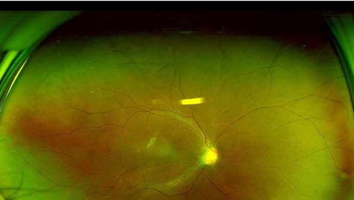
Fundus Explorer Pro
Photograph the retinal findings described here with the phone already in your pocket — 22 D optics and built-in illumination in one handheld unit.
From Choroida — the team behind this siteVariants That Deserve Attention
A few peripheral lesions carry a higher risk and need to be recognized.
- Lattice degeneration with retinal holes or tears, especially in symptomatic patients (see lattice degeneration)
- Horseshoe tears with a mobile flap and vitreous traction (see horseshoe tears)
- Atrophic holes within lattice or in a patient with a symptomatic PVD (see atrophic retinal holes)
- Retinal dialysis after trauma
- Meridional folds with a tear at the posterior end, a rare finding that can lead to detachment
- Enclosed oral bays with an associated tear, which need careful inspection
Risk rises with symptoms, myopia, prior detachment in the fellow eye, and family history.
Distinguishing Benign From Pathologic
The following points help.
- Symmetry: benign variants are usually bilateral and mirror images
- Location: ora serrata pearls, bays, and folds follow the anatomy of the ora
- Association with vitreous traction: a tear has a flap or operculum and vitreous attached to its edge
- Symptoms: new flashes and floaters raise suspicion
- Change over time: a stable lesion documented for years is reassuring
Scleral depression gives a dynamic view.
A break appears as a red defect with sharp edges, while a fold or bay moves with the indenter and the retina remains continuous.
Drawing the lesion or photographing it with widefield imaging creates a baseline for later comparison.
Management
No treatment is needed for normal variants.
For asymptomatic lattice, atrophic holes, or small retinal breaks, the evidence does not support routine prophylactic laser, and most are observed.
- The natural history of asymptomatic retinal breaks is favorable, with a low rate of detachment in long-term follow-up
- Prophylaxis is considered for symptomatic tears, for fresh horseshoe tears with persistent traction, and for selected high-risk situations such as a fellow eye that has detached (see laser retinopexy)
- Patients must be told about warning symptoms and given clear instructions to return promptly
A Cochrane review found insufficient evidence that prophylaxis of asymptomatic breaks and lattice prevents detachment.
Decisions should be individualized, and the reasoning documented.
Documenting Peripheral Findings
A reliable record makes later comparison possible.
- Draw the lesion on a fundus chart, using the standard color code, with its clock-hour position and distance from the ora
- Photograph with widefield imaging when available
- Note whether the lesion was found in one eye or both, and whether the vitreous was attached
- Record symptoms at the time of examination, including the date of onset
- State the reason for observing or treating, so that later examiners understand the decision
Patients also benefit from a short written note explaining that the findings are normal variants and listing the warning symptoms.
Teaching Points for Trainees
- Learn the normal anatomy of the ora serrata on scleral depression before judging pathology
- Examine both eyes, and compare
- Describe the lesion by position in clock hours and distance from the ora
- When in doubt, examine again after a few weeks and compare the drawings or photographs
- Do not treat a lesion simply because it looks unfamiliar
Good technique and repeated exposure make normal variants recognizable.


Document what you see
Two smartphone imaging tools built for everyday clinic use — one for the slit lamp, one for the fundus.
From Choroida — the team behind this siteReferences
- Byer NE. The natural history of asymptomatic retinal breaks. Ophthalmology. 1982;89:1033-1039.
- Byer NE. Long-term natural history of lattice degeneration of the retina. Ophthalmology. 1989;96:1396-1401.
- Straatsma BR, Zeegen PD, Foos RY, Feman SS, Shabo AL. Lattice degeneration of the retina. Trans Am Acad Ophthalmol Otolaryngol. 1974;78:OP87-OP113.
- Flaxel CJ, Adelman RA, Bailey ST, et al. Posterior vitreous detachment, retinal breaks, and lattice degeneration Preferred Practice Pattern. Ophthalmology. 2020;127:P146-P181.
- Wilkinson CP. Interventions for asymptomatic retinal breaks and lattice degeneration for preventing retinal detachment. Cochrane Database Syst Rev. 2014;(9):CD003170.
Test yourself
A few questions straight from this article.
-
What are peripheral retinal variants?
Peripheral retinal variants are normal or benign findings at the retinal periphery that resemble tears, holes or degenerations. Recognising them avoids unnecessary laser and unnecessary referral. -
What is an oral bay?
Oral bays are indentations of the pars plana epithelium into the retina, lying between dentate processes. When surrounded by retina they become enclosed oral bays, which can look like round holes. -
What is a meridional fold?
Meridional folds are radial ridges of redundant retina, usually lined up with a dentate process. A meridional complex combines a fold with a dentate or ciliary process. -
Where are most ora serrata variants found, and how are they distributed?
Most ora serrata variants are found in the nasal periphery and are often bilateral and symmetrical. They become more prominent on scleral depression. -
What is the appearance and significance of pavingstone degeneration?
Pavingstone degeneration appears as discrete yellow-white patches of chorioretinal atrophy between the ora and the equator, most often inferiorly. The retina over the patches is thinned but attached, and prophylactic treatment is not needed. -
Peripheral cystoid degeneration can coalesce to produce which condition?
Small intraretinal cysts at the ora are present in most older adults and are benign, but they may coalesce to form degenerative retinoschisis. -
On scleral depression, what distinguishes a retinal break from a meridional fold or oral bay?
A break appears as a red defect with sharp edges, while a fold or bay moves with the indenter and the retina remains continuous over it. Scleral depression gives this dynamic view. -
What does the evidence show about prophylactic laser for asymptomatic lattice degeneration and retinal breaks?
For asymptomatic lattice, atrophic holes or small breaks the evidence does not support routine prophylactic laser, and a Cochrane review found insufficient evidence that prophylaxis prevents detachment. Most such lesions are observed. -
In which situation is prophylactic treatment of a peripheral retinal break considered?
Prophylaxis is considered for symptomatic tears, for fresh horseshoe tears with persistent traction, and for selected high-risk situations such as a fellow eye that has detached. Patients must also be told the warning symptoms. -
How should a peripheral retinal lesion be recorded so that later comparison is possible?
The lesion is drawn on a fundus chart with its clock-hour position and distance from the ora, photographed with widefield imaging where available, and noted as unilateral or bilateral with the state of the vitreous.

Writes and reviews the clinical articles on Educate. Every post is checked against peer-reviewed sources before it goes live.


