
Uveitis masquerade syndromes are conditions that look like intraocular inflammation but are caused by something else, most often a malignancy.
The consequences of the wrong label are serious, because steroid treatment can mask a lymphoma for months while it spreads to the brain.
A few clinical clues, and the willingness to question the diagnosis when the course is atypical, reduce that risk.
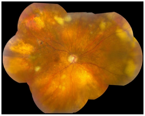
What Are Uveitis Masquerade Syndromes?
Masquerade syndromes divide into neoplastic and non-neoplastic groups.
The term was used to describe a set of diseases in which patients present with cells in the vitreous or anterior chamber, and are treated for uveitis without response.
In referral series from uveitis centers, a small but consistent proportion of patients sent for uveitis turned out to have a masquerade syndrome, and a sizeable share of these were neoplastic.
Neoplastic Masquerade Syndromes
Primary Vitreoretinal Lymphoma
This is the classic and most important masquerader.
- Typically affects patients over 50
- Presents with painless vitritis and floaters, often bilateral but asymmetric
- The anterior chamber is quiet or has mild cells
- Sub-RPE and subretinal yellow-white infiltrates with a leopard-spot pattern on autofluorescence
- Partial response to steroids, with relapse after tapering
- CNS lymphoma is present or develops later in a large share of patients
A high index of suspicion is required (see intraocular lymphoma).
Leukemia
Leukemic infiltration produces pseudohypopyon, vitreous cells, retinal infiltrates, and hemorrhages, and it may be the first sign of relapse.
Metastatic Carcinoma
Choroidal metastases cause serous detachment and can be mistaken for posterior scleritis or VKH (see choroidal metastases).
Anterior segment metastases may produce a hypopyon-like layer.
Uveal Melanoma
Necrotic melanoma can cause inflammation, vitritis, and pseudohypopyon.
Paraneoplastic Disease
Bilateral diffuse uveal melanocytic proliferation and cancer-associated retinopathy may resemble posterior uveitis (see BDUMP).
Retinoblastoma
In children, retinoblastoma with pseudohypopyon or vitreous seeding can mimic endophthalmitis or uveitis (see retinoblastoma).
Any child with white pupil, pseudohypopyon, or unexplained inflammation requires ultrasound and examination under anesthesia.
Juvenile Xanthogranuloma
This may cause spontaneous hyphema and iris nodules, and it presents in young children (see juvenile xanthogranuloma).
Fundus Explorer Pro
Photograph the retinal findings described here with the phone already in your pocket — 22 D optics and built-in illumination in one handheld unit.
From Choroida — the team behind this siteNon-Neoplastic Masquerade Syndromes
- Rhegmatogenous retinal detachment with anterior vitreous pigment cells, and chronic detachment with secondary inflammation (see Schwartz-Matsuo syndrome)
- Retinitis pigmentosa, with vitreous cells and cystoid macular edema (see retinitis pigmentosa)
- Intraocular foreign body, especially retained metal, with chronic inflammation
- Ocular ischemic syndrome, with anterior chamber flare and cells (see ocular ischemic syndrome)
- Chronic endophthalmitis, including low-grade infection after surgery
- Pigment dispersion with anterior chamber pigment
- Drug-induced effects, such as rifabutin uveitis and bisphosphonate-related inflammation
- Dry eye and ocular surface disease in mild cases
- Vitreous hemorrhage with red cells that resemble inflammatory cells
Clues That Suggest Masquerade
- Age over 50 with first-time unexplained vitritis
- Poor or incomplete response to corticosteroids, or a response followed by relapse
- Absence of anterior segment inflammation with dense vitritis
- Sub-RPE infiltrates, retinal masses, or a leopard-spot pattern
- A child with white pupil or pseudohypopyon
- Unilateral disease that progresses despite treatment
- Neurologic symptoms in a patient with vitritis
- Known cancer or a history of systemic malignancy
- Atypical retinal vasculitis or an unusual pattern of leakage
Revisiting the diagnosis in any patient who does not follow the expected course is the safest habit.
Investigations
Imaging
- OCT shows sub-RPE deposits and retinal infiltrates in lymphoma
- Autofluorescence shows the leopard-spot pattern
- Fluorescein angiography shows characteristic granular hypofluorescent lesions
- B-scan ultrasonography and ultrasound biomicroscopy show tumors, foreign bodies, and detachments
- MRI of the brain with contrast and lumbar puncture when CNS lymphoma is suspected
Vitreous and Aqueous Sampling
Diagnostic vitrectomy is the standard test when lymphoma is suspected.
- Cytology shows large atypical lymphoid cells, but cells are fragile and results depend on the handling of the specimen
- Flow cytometry and immunohistochemistry improve yield
- The ratio of interleukin-10 to interleukin-6 is elevated in lymphoma, and a ratio above one supports the diagnosis
- MYD88 L265P mutation is found in a large proportion of vitreoretinal lymphomas, and PCR testing increases diagnostic yield
Samples should be sent quickly to an experienced pathology laboratory, and steroids should be stopped for a period before biopsy when clinically safe, since they reduce diagnostic yield.
Management
Treatment follows the cause.
- Primary vitreoretinal lymphoma is treated with intravitreal methotrexate or rituximab, local radiotherapy, and systemic chemotherapy when CNS involvement is present, and management is shared with an oncologist
- Metastatic and leukemic disease need systemic treatment and radiotherapy
- Retinoblastoma needs urgent pediatric ocular oncology care
- Foreign bodies require removal
- Retinal detachments are repaired surgically
- Drug-induced uveitis improves after the drug is stopped
Early diagnosis affects survival in lymphoma and cancers.
Illustrative Presentations
- A 68-year-old with painless floaters in both eyes for eight months, partial improvement on oral prednisone, and yellow-white subretinal lesions: primary vitreoretinal lymphoma until proven otherwise
- A 45-year-old with pigment cells in the vitreous and mild inflammation in one eye, later found to have a retinal detachment: a detachment mimicking uveitis
- A young adult with night blindness, vitreous cells, and cystoid macular edema: retinitis pigmentosa
- A 3-year-old with a red eye, pseudohypopyon, and a white pupil: retinoblastoma until proven otherwise
Each case shows the same lesson, which is to look beyond the label of uveitis when the details do not fit.
Practical Message
Every patient with uveitis should be asked whether the pattern fits a known uveitis entity, whether the age is typical, and whether the response to treatment is what would be expected.
When the answers are unsatisfactory, a vitreous biopsy or a second opinion from a uveitis specialist is worth the delay.



Document what you see
Two smartphone imaging tools built for everyday clinic use — one for the slit lamp, one for the fundus.
From Choroida — the team behind this siteReferences
- Rothova A, Ooijman F, Kerkhoff F, Van Der Lelij A, Lokhorst HM. Uveitis masquerade syndromes. Ophthalmology. 2001;108:386-399.
- Chan CC, Rubenstein JL, Coupland SE, et al. Primary vitreoretinal lymphoma: a report from an International Primary Central Nervous System Lymphoma Collaborative Group symposium. Oncologist. 2011;16:1589-1599.
- Davis JL. Intraocular lymphoma: a clinical perspective. Eye (Lond). 2013;27:153-162.
- Bonzheim I, Giese S, Deuter C, et al. High frequency of MYD88 mutations in vitreoretinal B-cell lymphoma: a valuable tool to improve diagnostic yield of vitreous aspirates. Blood. 2015;126:76-79.
- Chan CC, Sen HN. Current concepts in diagnosing and managing primary vitreoretinal (intraocular) lymphoma. Discov Med. 2013;15:93-100.
