
CASE REPORT
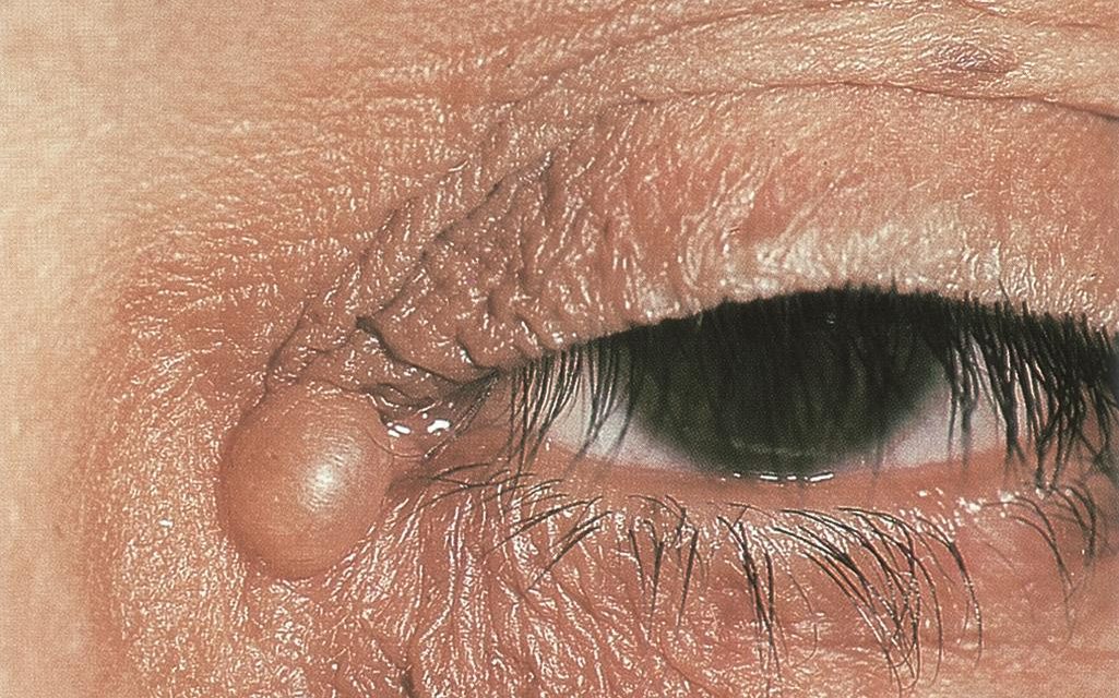
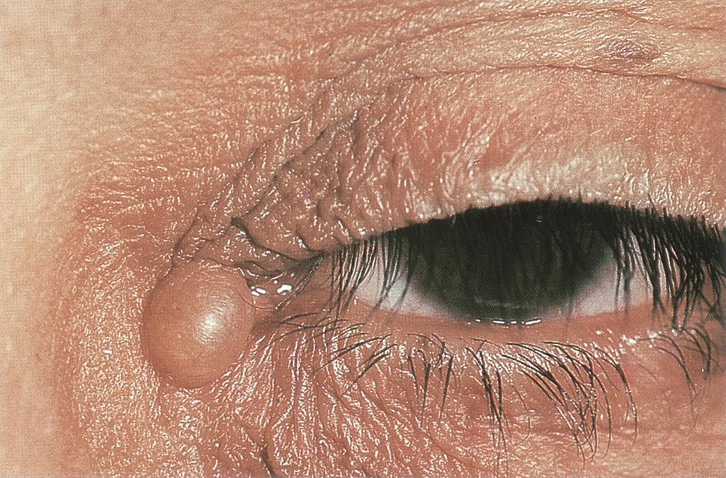
A 75-year-old male patient presented with small, translucent, cystic papules which were filled with a watery fluid, which had been present for five years and he was asymptomatic.
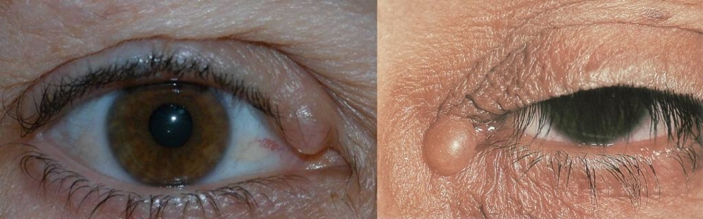
On local examination, the largest cyst measured 2×2 cm. The patient underwent, under local anesthesia, an excisional biopsy of the mass, and the specimen was subjected to a histopathological examination.
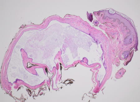
The gross examination of this specimen showed a grey-white cyst that measured 1.5×1 cm. The histopathologic examination of the paraffin sections which were stained with hematoxylin-eosin revealed the presence of a cyst in the dermis.
It was lined by an inner layer of a secretory columnar epithelium, which lay above an outer myoepithelial cell layer. At places, the secretory epithelium showed decapitation, which suggested an apocrine hidrocystoma.
Cyst in the dermis is lined by an inner layer of secretory columnar epithelium, which lies above an outer myoepithelial cell layer. At places, secretory epithelium shows decapitation, thus suggesting apocrine hidrocystoma
Apocrine Hidrocystoma entity
Apocrine Hidrocystomas are benign cystic tumors and are thought to be an adenomatous cystic proliferation of apocrine sweat glands.

These are frequently found in the head and neck region as apocrine glands are found in higher concentration in these areas, these lesions can be presented on the eyebrows, upper and lower eyelids, and medial or lateral canthi.
Apocrine Hidrocystomas do not follow any inheritance pattern and have been observed in middle-aged and elderly people with no predilection for sex and race.
These are usually asymptomatic or may have a symptom cosmetically unappealing for the patient. Others mild symptoms of itching, eye irritation, and foreign body sensation can also be present.
These are mobile upon palpation and are presented as a dome–shaped, translucent, or blue-gray-purple cystic nodule with a size ranging from 3-15mm.
Apocrine Hidrocystoma MANAGEMENT
- Complete surgical excision along with the walls of cysts is the best treatment option for apocrine hidrocystomas.
- Needle puncture is another method. When apocrine hidrocystomas are incised a brownish fluid is released. However, this treatment provides a temporary recovery, and the recurrence chances are greater.
- Removal with trichloroacetic acid is another best treatment plan for apocrine lesions. In this method after local anesthesia first, the cyst content is aspirated with an empty syringe and then refilled with another syringe containing 20% TCA. Left it for 1 minute in the cyst and then aspirated and washed with distilled water.
- Intradermal injections of low-dose botulinum toxin A around the cyst are also being performed nowadays.
HOW TO TAKE SLIT-LAMP EXAM IMAGES WITH A SMARTPHONE?
Smartphone slit-lamp photography is the new advancement in the field of science and technology in which photographs of the desired slit-lamp finding can be taken with smartphones by using the slit-lamp adapters.
Slit-lamp Smartphone photography
REFERENCES
- Hafsi W, Badri T, Shah F. Apocrine Hidrocystoma. [Updated 2021 Sep 9]. In: StatPearls [Internet]. Treasure Island (FL): StatPearls Publishing; 2021 Jan.
- Mehregan AH. Apocrine Cystadenoma; A Clinicopathologic Study With Special Reference To The Pigmented Variety. Arch Dermatol. 1964;90:274–9.
- Magdaleno-Tapial J, Valenzuela-Oñate C, Martínez-Doménech Á, García-Legaz-Martínez M, Martínez-Aparicio A, Alegre-de Miquel V, Pérez-Ferriols A. Apocrine hidrocystoma on the nipple: the first report in this unusual location. Dermatol Online J. 2019 Oct 15;25(10)
- Nam JH, Lee GY, Kim WS, Kim KJ. Eccrine hidrocystoma in a child: an atypical presentation. Ann Dermatol. 2010 Feb;22(1):69-72.
- Birkenbeuel J, Goshtasbi K, Mahboubi H, Djalilian HR. Recurrent apocrine hidrocystoma of the external auditory canal. Am J Otolaryngol. 2019 Mar – Apr;40(2):312-313.
- Sarabi K, Khachemoune A. Hidrocystomas–a brief review. MedGenMed. 2006 Sep 06;8(3):57.
Slit-lamp Smartphone photography

RETINAL IMAGING BY YOUR SMARTPHONE
Thanks