
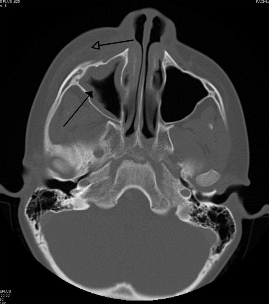
Orbital cellulitis is infection posterior to the orbital septum, involving the fat and muscles within the bony orbit, and it is the diagnosis that must be excluded confidently in every child or adult presenting with a swollen, red eyelid.
The distinction from preseptal cellulitis, infection confined anterior to the septum, is not academic. Orbital cellulitis carries genuine risk of vision loss, cavernous sinus thrombosis, and intracranial spread, while preseptal disease, though it needs treatment, does not carry that same risk profile.
Anatomy and Pathophysiology
The orbital septum is a fibrous sheet that separates the eyelid and periorbital soft tissue from the orbital contents proper. Infection anterior to the septum, preseptal cellulitis, is generally milder and typically arises from local skin trauma, an insect bite, or spread from a chalazion or hordeolum.
Infection posterior to the septum involves the orbital fat and extraocular muscles directly and behaves as a genuinely different, more dangerous disease.
The great majority of orbital cellulitis in both children and adults arises from contiguous spread of sinusitis, most often ethmoid, given the thin lamina papyracea separating the ethmoid sinuses from the orbit. Direct extension through this thin bony wall, or via the valveless ophthalmic veins that drain both regions, allows a sinus infection to become an orbital one over a matter of days.
Less commonly, infection follows orbital trauma, dental infection, dacryocystitis, or, in adults, spread from odontogenic sources through the maxillary sinus.
Clinical Presentation
The features that separate orbital from preseptal disease are worth checking specifically and explicitly in every patient with periorbital swelling, because relying on overall clinical impression alone is where the diagnosis gets missed.
Proptosis, pain with eye movement, and restricted or painful ocular motility all point to orbital involvement. Reduced visual acuity or a relative afferent pupillary defect are red flags for optic nerve compromise and demand urgent action rather than routine follow-up.
Chemosis can occur in either preseptal or orbital disease and is therefore less discriminating on its own, but it tends to be more marked with orbital involvement. Fever and systemic illness are more common with orbital disease, though their absence does not exclude it, particularly early in the course or in a patient already on oral antibiotics for presumed sinusitis.
Children, in particular, may present with a preceding history of upper respiratory infection or sinusitis, and the periorbital swelling can develop rapidly over 24 to 48 hours. Any child with marked proptosis, pain on eye movement, or reduced vision needs urgent assessment rather than a trial of oral antibiotics with review in a few days.
All-fit Slit-Lamp Adapter
Record and share exactly what you see at the slit lamp. One adapter fits any slit lamp or surgical microscope — and any smartphone.
From Choroida — the team behind this siteDifferential Diagnosis
- Preseptal cellulitis, distinguished by the absence of proptosis, painful or restricted motility, and visual compromise
- Orbital inflammatory disease or idiopathic orbital inflammation, which can mimic infection closely and sometimes only declares itself with a lack of response to antibiotics or with imaging and inflammatory marker patterns that do not fit infection
- Thyroid eye disease, generally more gradual in onset and typically bilateral, without the acute pain and fever of cellulitis
- Orbital or periorbital tumour, particularly rhabdomyosarcoma in a child, which can present with rapid proptosis and should be considered if the clinical course does not follow the expected trajectory for infection
- Allergic or contact dermatitis causing eyelid swelling, generally itchy rather than painful and without proptosis or motility restriction
Diagnostic Evaluation
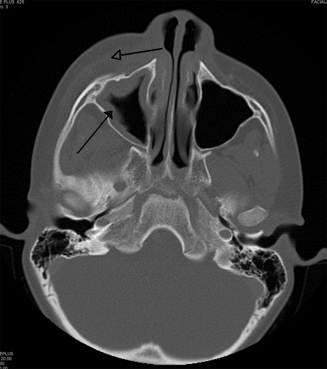
Contrast-enhanced CT of the orbits and sinuses is the primary imaging study, both to confirm orbital involvement and to identify the underlying sinusitis and any subperiosteal or orbital abscess that would change management.
MRI is preferred when there is concern for intracranial extension or cavernous sinus involvement, given its superior soft tissue resolution for those structures.
Blood cultures should be obtained, particularly in patients unwell enough to require admission, though yield is often low. Where a subperiosteal or orbital abscess requires surgical drainage, culture of the purulent material provides the most useful microbiological guidance for narrowing therapy.
Management
Orbital cellulitis is a medical and surgical emergency requiring hospital admission and intravenous broad-spectrum antibiotics covering the likely pathogens, principally Staphylococcus aureus, including consideration of MRSA depending on local resistance patterns, streptococcal species, and, especially in sinusitis-associated disease, anaerobes.
Specific antibiotic choice should follow local microbiology guidance and be adjusted once culture results are available.
Surgical drainage is indicated for a large or medially located subperiosteal abscess, for any abscess causing significant proptosis or threatening the optic nerve, and for cases failing to improve on appropriate intravenous antibiotics within 24 to 48 hours.
Smaller, well-localised abscesses in an otherwise improving patient are sometimes managed with continued medical therapy and close monitoring, so the decision to operate is not automatic on imaging alone but depends on size, location, and clinical trajectory.
Vision, pupillary response, and eye movements should be monitored closely throughout treatment, since deterioration on appropriate antibiotics, or any sign of worsening optic nerve function, is an indication for urgent repeat imaging and surgical reassessment rather than simply extending the antibiotic course.
Complications
- Subperiosteal or orbital abscess formation, present in a meaningful proportion of cases at diagnosis
- Optic neuropathy from direct compression or ischaemia, with risk of permanent vision loss if not addressed promptly
- Cavernous sinus thrombosis, a rare but life-threatening extension along the valveless orbital venous system
- Intracranial extension, including meningitis, epidural or subdural abscess, and cerebral abscess
- Central retinal artery or vein occlusion from severe orbital pressure or associated inflammation
Cavernous sinus thrombosis in particular should be suspected if bilateral orbital signs develop, given the direct venous communication between the two orbits through the cavernous sinus, and this finding should prompt urgent neuroimaging and neurology or neurosurgery involvement.
Prognosis
With prompt diagnosis and appropriate treatment, most patients recover fully without permanent visual or neurological sequelae.
Outcome is closely tied to how quickly treatment begins relative to symptom onset, and to whether optic nerve compromise develops before intervention. Delayed presentation or delayed recognition, particularly cases initially misdiagnosed and treated as preseptal disease, carries a meaningfully higher risk of complications.
Children generally have good outcomes with timely treatment, though severe cases can still result in permanent visual loss or, rarely, intracranial complications, which is the underlying reason this diagnosis is treated with the urgency it is.


Document what you see
Two smartphone imaging tools built for everyday clinic use — one for the slit lamp, one for the fundus.
From Choroida — the team behind this siteReferences
- Chandler JR, Langenbrunner DJ, Stevens ER. The pathogenesis of orbital complications in acute sinusitis. Laryngoscope. 1970.
- Bedwell J, Bauman NM. Management of pediatric orbital cellulitis and abscess. Current Opinion in Otolaryngology & Head and Neck Surgery. 2011.
- Chaudhry IA, Al-Rashed W, Arat YO. The hot orbit: orbital cellulitis. Middle East African Journal of Ophthalmology. 2012.
- Orbital Cellulitis. EyeWiki, American Academy of Ophthalmology.
- Orbital Cellulitis. StatPearls, NCBI Bookshelf.
Test yourself
A few questions straight from this article.
-
Orbital cellulitis is defined by infection in which anatomical location?
The orbital septum is the dividing line: infection behind it involves the orbital fat and extraocular muscles directly. Preseptal cellulitis, confined anterior to the septum, is a milder and genuinely different disease. -
What is the source of the great majority of orbital cellulitis cases?
The thin lamina papyracea separates the ethmoid sinuses from the orbit, allowing direct extension. Valveless ophthalmic veins draining both regions provide a second route, so sinus infection can become orbital within days. -
Which combination of findings points to orbital rather than preseptal cellulitis?
Proptosis, pain on eye movement and restricted motility all indicate postseptal involvement. These features should be checked explicitly in every patient with periorbital swelling, since overall impression alone is where the diagnosis gets missed. -
Which finding in orbital cellulitis is a red flag demanding urgent action rather than routine follow-up?
Reduced visual acuity or a relative afferent pupillary defect signals optic nerve compromise. Vision, pupillary response and eye movements need close monitoring throughout treatment, since deterioration mandates urgent reassessment. -
Which imaging study is the primary investigation in suspected orbital cellulitis?
Contrast CT confirms orbital involvement, identifies the underlying sinusitis, and shows any subperiosteal or orbital abscess that would change management. It is the study that drives the surgical decision. -
When is MRI preferred over CT in orbital cellulitis?
MRI offers superior soft tissue resolution for intracranial structures and the cavernous sinus. For the orbit and sinuses themselves, contrast-enhanced CT remains the primary study. -
Which pathogens must empirical intravenous therapy for orbital cellulitis cover?
Broad-spectrum intravenous cover targets Staphylococcus aureus, including MRSA where local resistance warrants, streptococcal species, and anaerobes in sinusitis-associated disease. Choice follows local microbiology guidance and is narrowed once cultures return. -
Failure to improve on appropriate intravenous antibiotics over what period is an indication for surgical drainage?
Lack of improvement within 24 to 48 hours on appropriate intravenous antibiotics is a drainage indication, alongside a large or medial subperiosteal abscess or one threatening the optic nerve. Smaller abscesses in an improving patient may be watched. -
The development of bilateral orbital signs during orbital cellulitis should raise suspicion of which complication?
The two orbits communicate directly through the cavernous sinus by way of the valveless venous system, so bilateral signs suggest thrombosis there. This warrants urgent neuroimaging and neurology or neurosurgery involvement. -
Which factor most strongly determines outcome in orbital cellulitis?
Outcome tracks the delay between symptom onset and treatment, and whether optic nerve compromise develops first. Cases initially misdiagnosed and treated as preseptal disease carry a meaningfully higher risk of complications.

Writes and reviews the clinical articles on Educate. Every post is checked against peer-reviewed sources before it goes live.


