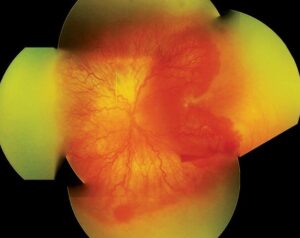
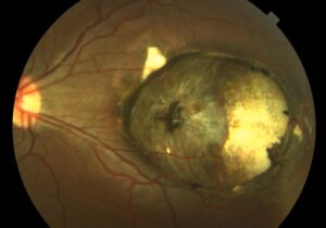
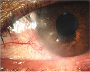
A photograph taken with a flash can occasionally reveal a cancer before any doctor does.
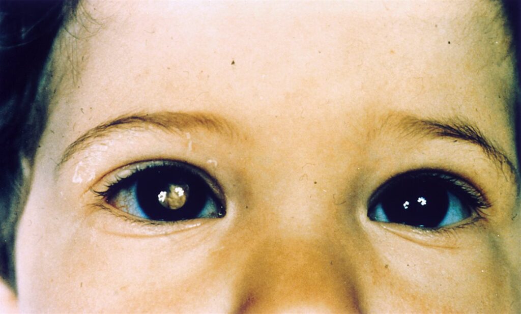
Retinoblastoma is the most common primary intraocular malignancy of childhood, and it most often first shows itself as an abnormal white reflection in a photo or in ambient light.
That white reflex — leukocoria — replaces the normal red-eye reflection in the affected eye.
Parents frequently notice it before any clinician does, which makes public awareness of this single sign a genuine part of early detection.
Survival is excellent when caught early, but delay allows both vision loss and, in advanced cases, life-threatening spread.
Recognizing leukocoria, understanding the urgency it demands, and knowing the modern treatment landscape are essential for anyone who examines children’s eyes.
What Is Retinoblastoma?
Retinoblastoma is a malignant tumor arising from immature retinal cells, almost always presenting in early childhood.
Key features include:
- The most common primary intraocular cancer in children, though still rare overall
- Can be unilateral (more common) or bilateral, with bilateral disease presenting at a younger age
- Arises from a mutation in the RB1 tumor suppressor gene, either inherited (germline) or sporadic (somatic)
- Highly curable when detected early, with survival rates exceeding 95% in high-resource settings
The distinction between heritable and non-heritable disease matters enormously, since heritable cases carry lifelong risk of second cancers and have implications for siblings and future children.
Epidemiology
Retinoblastoma is rare in absolute terms but is the most common childhood eye cancer worldwide.
- Most cases are diagnosed before age 2, and the great majority before age 5
- Bilateral cases present earlier, often in infancy, and are essentially always heritable
- Unilateral cases are usually sporadic, though a minority still carry a germline mutation
- Global outcomes vary sharply by resource setting — survival is excellent where early detection and treatment access are strong, and far worse where diagnosis is delayed
The age pattern itself is diagnostically useful: a very young infant with bilateral leukocoria should raise immediate suspicion for heritable disease.
Fundus Explorer Pro
Photograph the retinal findings described here with the phone already in your pocket — 22 D optics and built-in illumination in one handheld unit.
From Choroida — the team behind this sitePathophysiology
Retinoblastoma follows the classic “two-hit” model of tumor suppressor gene inactivation.
- Both copies of the RB1 gene must be inactivated for a retinal cell to become malignant
- In heritable disease, the first mutation is present in every cell from birth, so only one further somatic mutation is needed — explaining earlier onset and frequent bilateral, multifocal tumors
- In non-heritable disease, both mutations must occur independently within the same retinal cell, a rarer event that explains later onset and typically unilateral, single-focus disease
- Loss of functional RB1 protein removes a key brake on cell cycle progression, allowing uncontrolled proliferation of retinal progenitor cells
- Tumor growth within the eye eventually obstructs the visual axis or fills the vitreous cavity, producing the white pupillary reflex
Because heritable disease carries a germline mutation in every cell, survivors also carry a lifelong elevated risk of other cancers, particularly osteosarcoma and other sarcomas.
Risk Factors
Genetic Risk Factors
- A germline RB1 mutation, whether inherited from an affected parent or arising as a new mutation
- A family history of retinoblastoma in a parent or sibling
Other Considerations
- Most cases occur with no family history at all, reflecting sporadic somatic mutation
- No consistently established environmental or maternal risk factor has been identified for sporadic disease
Genetic counseling is relevant for every family with a diagnosed child, since it directly affects screening recommendations for siblings and future children.
Clinical Presentation
Symptoms and Signs Noticed by Family
- Leukocoria — an abnormal white pupillary reflection, often first noticed in flash photographs
- Strabismus (ocular misalignment), when the tumor involves the macula and disrupts central fixation
- Less commonly, a red, painful eye from secondary glaucoma or inflammation in advanced disease
- Rarely, proptosis or orbital signs in very advanced, extraocular extension
Examination Findings
- A white or yellow-white pupillary reflex replacing the normal red reflex, often asymmetric between the two eyes
- One or more creamy white to pink retinal masses on dilated fundus examination, sometimes with visible calcification
- Vitreous or subretinal seeding in more advanced disease
- Absent red reflex on direct ophthalmoscopy or photographic screening — a simple, low-tech finding with high diagnostic value
A red reflex test takes seconds and is one of the highest-yield screening maneuvers in all of pediatric eye care — an asymmetric or absent reflex should never be dismissed.
Diagnostic Evaluation
Examination Under Anesthesia
- Detailed dilated fundus examination, typically performed under anesthesia in young children, to map every tumor focus, its size, and location
- Careful documentation used for staging and to guide treatment planning
Imaging
- Ocular ultrasound to detect calcification within the tumor, a supportive diagnostic feature
- MRI of the orbits and brain to assess for optic nerve invasion, extraocular extension, and, in bilateral cases, pineal involvement (trilateral retinoblastoma)
- CT is generally avoided given radiation exposure risk, particularly in children with a germline RB1 mutation
Genetic Testing
- RB1 genetic testing for the child and, where indicated, family members to determine heritable versus sporadic status
- Results guide screening intensity for at-risk siblings and future children
Biopsy of the tumor itself is avoided given the risk of seeding tumor cells outside the eye — diagnosis relies on examination and imaging rather than tissue sampling.
Differential Diagnosis
Conditions that can mimic retinoblastoma’s leukocoria include:
- Persistent fetal vasculature (persistent hyperplastic primary vitreous) — typically unilateral with an associated small, malformed eye
- Coats disease — retinal telangiectasia with exudation, occurring in older children and typically boys
- Retinopathy of prematurity — history of prematurity and characteristic peripheral findings
- Toxocariasis — history of exposure, often with a peripheral granuloma rather than a central mass
- Congenital cataract — an opacity within the lens itself rather than a retinal mass
Because several of these mimics are benign, prompt specialist evaluation is essential to distinguish them from a genuine malignancy without unnecessary delay in either direction.
Management
Eye-Preserving (Globe-Salvage) Therapy
- Intravenous, intra-arterial, or intravitreal chemotherapy to reduce tumor burden while preserving the eye and useful vision when possible
- Focal treatments — laser photocoagulation or cryotherapy — for smaller tumors, often combined with chemotherapy
- Intra-arterial chemotherapy delivered directly to the ophthalmic artery has become an important option for eyes that previously would have required enucleation
Enucleation
- Reserved for eyes with advanced, extensive disease with no reasonable chance of useful vision, or where the risk of extraocular spread is high
- Remains a definitive, sight-sacrificing but life-preserving option when disease extent precludes globe-salvage therapy
Systemic and Long-Term Care
- Systemic chemotherapy or radiotherapy for extraocular or metastatic disease
- Lifelong surveillance for heritable disease survivors, given elevated risk of second malignancies, particularly osteosarcoma
- Genetic counseling and screening protocols for at-risk siblings and future offspring
Treatment planning is highly individualized, balancing tumor control, eye and vision preservation, and long-term survivorship risk in every decision.
Prognosis
Prognosis has improved dramatically with modern treatment, though outcomes still depend heavily on timing and access to care.
- Survival exceeds 95% in high-resource settings with early diagnosis and treatment
- Outcomes are considerably worse in settings where diagnosis and treatment access are delayed
- Heritable disease survivors face an elevated lifetime risk of second cancers, requiring long-term oncologic follow-up beyond the eye itself
The single biggest determinant of outcome remains how early the disease is recognized — which puts real weight on a simple, familiar sign that any parent or clinician can learn to notice.


Document what you see
Two smartphone imaging tools built for everyday clinic use — one for the slit lamp, one for the fundus.
From Choroida — the team behind this siteReferences
- Dimaras H, Corson TW, Cobrinik D, et al. Retinoblastoma. Nature Reviews Disease Primers. 2015.
- Fabian ID, Abdallah E, Abdullahi SU, et al. Global Retinoblastoma Presentation and Analysis by National Income Level. JAMA Oncology. 2020.
- Shields CL, Shields JA. Retinoblastoma management: advances in enucleation, intravenous chemoreduction, and intra-arterial chemotherapy. Current Opinion in Ophthalmology. 2010.
- Abramson DH, Shields CL, Munier FL, Chantada GL. Treatment of Retinoblastoma in 2015: Agreement and Disagreement. JAMA Ophthalmology. 2015.
- Retinoblastoma. EyeWiki, American Academy of Ophthalmology.
Test yourself
A few questions straight from this article.
-
Retinoblastoma is best described as which type of tumour?
Retinoblastoma is a malignant tumour arising from immature retinal cells and is the most common primary intraocular cancer in children, though still rare overall. -
Inactivation of which gene underlies retinoblastoma?
Both copies of the RB1 tumour suppressor gene must be inactivated for a retinal cell to become malignant; loss of RB1 protein removes a key brake on cell cycle progression. -
What does bilateral retinoblastoma presenting in an infant imply?
Bilateral disease presents earlier, often in infancy, and is essentially always heritable, because the first RB1 mutation is already present in every cell from birth. -
What is the survival rate for retinoblastoma diagnosed and treated early in high-resource settings?
Retinoblastoma is highly curable when detected early, with survival exceeding 95% in high-resource settings; outcomes are considerably worse where diagnosis and treatment access are delayed. -
Which presenting sign of retinoblastoma is most often noticed first by a parent?
Leukocoria replaces the normal red-eye reflection and is frequently first spotted in a flash photograph, which is why public awareness of this one sign contributes to early detection. -
Which ocular ultrasound finding supports a diagnosis of retinoblastoma?
Ocular ultrasound is used to detect calcification within the tumour, a supportive diagnostic feature that also helps separate retinoblastoma from benign causes of leukocoria. -
Why is CT generally avoided in the workup of retinoblastoma?
CT is avoided because of radiation exposure, particularly in children carrying a germline RB1 mutation. MRI of the orbits and brain is preferred to assess optic nerve invasion and pineal involvement. -
Why is biopsy avoided when diagnosing retinoblastoma?
Sampling the tumour risks seeding malignant cells outside the eye, so diagnosis rests on examination under anaesthesia and imaging rather than on tissue. -
Which globe-salvage treatment for retinoblastoma is delivered directly into the ophthalmic artery?
Intra-arterial chemotherapy delivered into the ophthalmic artery has become an important option for eyes that previously would have required enucleation. -
Survivors of heritable retinoblastoma are at particular lifelong risk of which second malignancy?
Because the germline RB1 mutation is present in every cell, survivors of heritable disease carry a raised lifetime risk of second cancers, particularly osteosarcoma and other sarcomas.

Writes and reviews the clinical articles on Educate. Every post is checked against peer-reviewed sources before it goes live.