
Epiblepharon is a common congenital eyelid anomaly in which an extra horizontal fold of skin and underlying orbicularis muscle pushes the eyelashes into a vertical position against the cornea or conjunctiva.
While it can look alarmingly similar to entropion on first glance, the two conditions have different mechanisms, different natural histories, and often different management, which makes correctly distinguishing them clinically important rather than a mere academic exercise for the examining clinician.
Epiblepharon vs. Entropion
The key structural difference is that in epiblepharon the eyelid margin itself remains in its normal anatomic position — it is only the redundant skin fold riding over the margin that redirects the lashes vertically — whereas true entropion involves actual inward rotation of the tarsal plate and lid margin itself.
This distinction explains why epiblepharon in children so often resolves spontaneously as the child’s face grows and the redundant fold is gradually pulled taut by normal facial development, a resolution pattern that true entropion does not share.
Because the underlying lid margin architecture is normal in epiblepharon, there is no structural abnormality that needs surgical correction unless the cornea is genuinely being irritated — the redundant skin fold itself, not the lid margin, is the only anatomic target if surgery is eventually needed.
Epidemiology
Epiblepharon is considerably more common in children of East Asian descent than in other populations, reflecting typical differences in facial and lid anatomy across ethnic groups.
It typically affects the lower eyelid, though upper lid involvement occurs as well, sometimes bilaterally and sometimes affecting all four lids in the same child.
It is usually noted in infancy or early childhood and, when the lower lid is involved, tends to improve as the nasal bridge and midface develop with growth, often resolving without any treatment by later childhood, which is worth explaining clearly to parents who may otherwise worry that nothing is being actively done about the finding.
All-fit Slit-Lamp Adapter
Record and share exactly what you see at the slit lamp. One adapter fits any slit lamp or surgical microscope — and any smartphone.
From Choroida — the team behind this siteClinical Presentation
Many affected children are entirely asymptomatic despite lashes visibly touching the ocular surface, because fine, soft, vellus-type infant and childhood lashes are often less irritating to the cornea than the coarser lashes of an adult with entropion-related trichiasis.
This is a genuinely important difference that shapes the threshold for intervention.
When symptomatic, children may show excessive tearing, blinking, or eye rubbing, or a parent may notice recurrent redness, though many cases are picked up incidentally on routine pediatric or ophthalmic exam rather than because of a specific complaint.
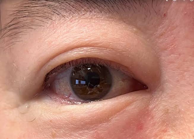
Exam Findings
- A horizontal fold of skin and muscle overriding the eyelid margin, most often the lower lid, pushing lashes into a vertical orientation
- The lid margin itself in normal anatomic position, distinguishing it from true entropion
- Variable degrees of corneal fluorescein staining depending on how much contact and irritation the lashes are actually causing
- Often bilateral, sometimes involving both upper and lower lids
Differential Diagnosis
- Congenital entropion — true inward rotation of the lid margin and tarsal plate, a distinct and much rarer diagnosis requiring different surgical correction
- Distichiasis — an accessory row of lashes emerging from meibomian gland orifices, a separate anatomic abnormality rather than a skin fold pushing normal lashes into contact with the eye
- Trichiasis from another cause — misdirected lashes without either lid margin rotation or an overriding skin fold
Management
Observation is appropriate for most children, when the cornea shows minimal or no staining, given the strong tendency toward spontaneous improvement with facial growth, especially in lower lid disease.
Lubrication can be used for mild irritation while awaiting natural resolution, and parents can be reassured that most cases do not require surgical intervention, though periodic follow-up remains worthwhile to confirm the expected trajectory of improvement is actually occurring.
Surgical correction is reserved for children with significant, persistent corneal staining, symptomatic irritation, or cases that fail to improve as expected with growth, typically involving excision of the redundant skin and muscle fold along with anchoring sutures to prevent recurrence — a different and generally simpler procedure than the lid-tightening or posterior lamellar surgery used for true entropion.
Upper lid epiblepharon and cases persisting into later childhood without improvement are more likely to require surgical correction than typical lower lid disease in a young child.
Recurrence after surgery is uncommon when the redundant skin and muscle are adequately addressed, and most children who do proceed to surgical correction have an excellent long-term outcome with resolution of the corneal irritation that prompted the intervention.
Why the Distinction From Entropion Matters
Because epiblepharon so often resolves on its own, defaulting to observation with monitoring — rather than reflexively pursuing surgery as might be more appropriate for entropion — spares many children an unnecessary procedure.
Conversely, mistaking true congenital entropion for epiblepharon and observing it risks allowing ongoing corneal irritation and potential scarring to continue in a condition that would not have resolved on its own the way epiblepharon typically does.
Careful documentation of the lid margin position at the initial exam — specifically noting whether the margin itself is rotated inward or simply overridden by a skin fold — is what allows this distinction to be made confidently and tracked reliably at follow-up visits.


Document what you see
Two smartphone imaging tools built for everyday clinic use — one for the slit lamp, one for the fundus.
From Choroida — the team behind this siteReferences
- Noda S, Hayasaka S, Setogawa T. Epiblepharon with inverted eyelashes in Japanese children. Graefe’s Archive for Clinical and Experimental Ophthalmology.
- Kwitko ML. Congenital malposition of the eyelids: entropion, epiblepharon, and euryblepharon. In: Pediatric Ophthalmology and Strabismus.
- American Academy of Ophthalmology. Basic and Clinical Science Course, Section 6: Pediatric Ophthalmology and Strabismus.
Test yourself
A few questions straight from this article.
-
In epiblepharon, what pushes the eyelashes into a vertical position against the ocular surface?
Epiblepharon is a congenital anomaly in which an extra horizontal fold of skin and underlying orbicularis rides over the lid, redirecting otherwise normal lashes vertically. -
Which structural feature separates epiblepharon from true entropion on examination?
In epiblepharon only the skin fold overrides the margin; true entropion involves actual inward rotation of the tarsal plate and lid margin. -
Why does lower lid epiblepharon in children so often resolve without any treatment?
As the nasal bridge and midface develop, the redundant fold is drawn taut and the lashes rotate away, a resolution pattern true entropion does not share. -
In which group is epiblepharon considerably more common?
Epiblepharon is considerably more common in children of East Asian descent, reflecting typical differences in facial and lid anatomy across populations. -
Why are many children with epiblepharon symptom-free despite lashes touching the eye?
Fine, soft infant and childhood lashes irritate the cornea far less than the coarser lashes of an adult with entropion-related trichiasis, which raises the threshold for intervention. -
Which examination finding best gauges how much the lashes are actually irritating the cornea in epiblepharon?
Corneal fluorescein staining varies with how much contact and irritation the lashes are causing, and it is the finding that drives the decision to observe or operate. -
Which feature defines distichiasis rather than epiblepharon?
Distichiasis is a separate anatomic abnormality, an extra lash row from the meibomian orifices, rather than a skin fold pushing normal lashes against the eye. -
What is appropriate management for most children with epiblepharon and minimal corneal staining?
Given the strong tendency to improve with facial growth, observation spares most children an unnecessary procedure, with lubrication for mild irritation and follow-up to confirm the expected improvement. -
What does surgical correction of epiblepharon typically involve?
Since the lid margin is normal, the redundant fold is the only anatomic target; excision with anchoring sutures to prevent recurrence is simpler than entropion surgery. -
Which presentations of epiblepharon are more likely to require surgical correction?
Typical lower lid disease in a young child tends to resolve with growth, whereas upper lid involvement and persistence without improvement more often come to surgery.
