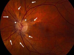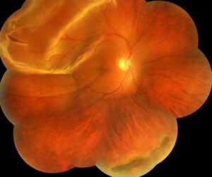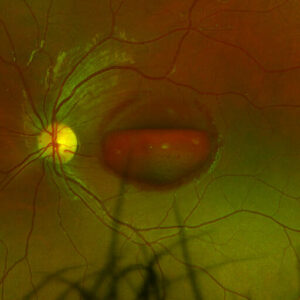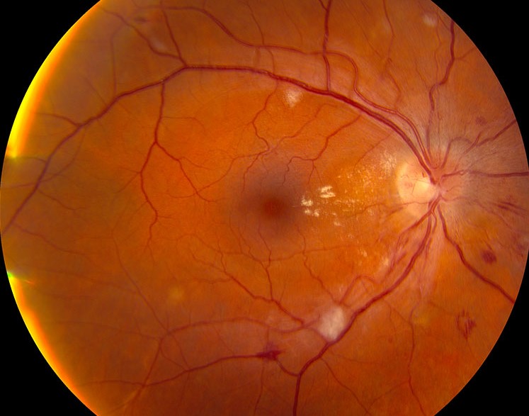
Blot hemorrhages are small, round, dark red bleeds that lie deep in the sensory retina.
They usually arise from the deep capillary network, so their shape reveals their depth.
However, shape alone does not name the disease.
Therefore, the examiner must also read their number, distribution and associated signs.
What Are Blot Hemorrhages?
Dot hemorrhages look tiny and pinpoint, while blot hemorrhages appear larger with slightly irregular edges.
Both lie mainly within the inner nuclear and outer plexiform layers.
No sharp biological boundary separates them, because the terms describe a size spectrum.
As a result, clinicians often record them together as dot and blot hemorrhages.
Why Blot Hemorrhages Look Round
In the middle retina, bipolar cells and Müller fibers run roughly perpendicular to the surface.
Blood therefore tracks along these vertical columns and spreads little sideways.
From above, the examiner sees the end of that column as a round spot.
In contrast, superficial bleeds follow the nerve fiber axons and form flame shapes instead (see retinal hemorrhage patterns).
Fundus Explorer Pro
Photograph the retinal findings described here with the phone already in your pocket — 22 D optics and built-in illumination in one handheld unit.
From Choroida — the team behind this siteFundus Examination Signs
First, use a dilated examination with a 90 D lens, then scan the periphery with indirect ophthalmoscopy.
Retinal vessels pass over blot hemorrhages, which helps confirm their deep position.
Also, red-free light makes small lesions stand out.
However, patients often report no symptoms unless the macula is involved or edema coexists.
Blot Hemorrhages on Imaging
On fluorescein angiography, blood blocks fluorescence and stays dark throughout the study.
Likewise, OCT typically shows a hyperreflective focus in the middle retina with shadowing beneath it.
Blot Hemorrhages in Diabetic Retinopathy
Diabetes damages capillaries through pericyte loss and basement membrane changes.
As a result, weakened walls leak and rupture, which produces deep bleeds in the posterior pole.
In addition, microaneurysms, hard exudates and cotton wool spots often accompany them.
Early disease may show only scattered lesions and normal vision.
Grading With the 4-2-1 Rule
First, count hemorrhages by quadrant, because extent carries prognostic weight.
Severe hemorrhages and microaneurysms in all four quadrants meet one criterion for severe nonproliferative disease.
Also, venous beading in two quadrants, or intraretinal microvascular abnormalities in one, qualifies.
As a result, eyes at this level face a substantially higher risk of proliferative disease.
Large Dark Blot Hemorrhages
Many retina specialists regard large, dark blots as a warning of nonperfused retina.
However, the association rests largely on clinical and angiographic observation, so treat it as a clue.
Then widefield angiography can map the nonperfused area directly.
Blot Hemorrhages in Vein Occlusion
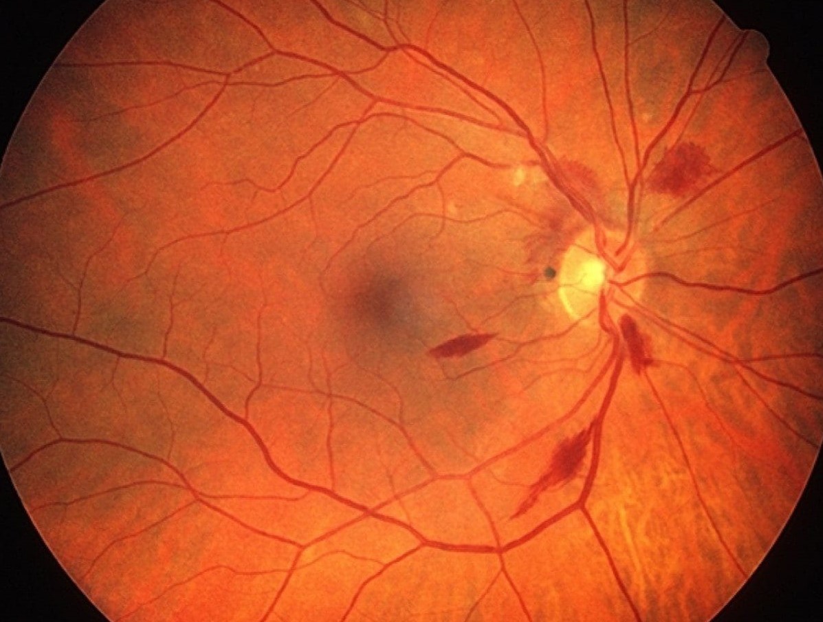
Central vein occlusion produces hemorrhages in all four quadrants, with dilated, tortuous veins.
Flame and deep hemorrhages usually appear together, and the disc may swell.
In contrast, branch occlusion confines the bleeding to one sector, which often respects the horizontal raphe.
Extensive deep hemorrhage with a relative afferent pupillary defect raises concern for ischemic central retinal vein occlusion.
Blot Hemorrhages in Ocular Ischemia
Carotid disease typically produces fewer blots, and they favor the mid-periphery.
Also, the veins look dilated and irregular, but they usually lack marked tortuosity.
Delayed choroidal filling and a prolonged arteriovenous time then support ocular ischemic syndrome.
Other Causes to Consider
Bilateral lesions without a typical diabetic pattern should prompt a wider search.
- For example, systemic hypertension
- Also, severe anemia or thrombocytopenia
- In addition, leukemia and hyperviscosity states
- Then radiation retinopathy
- Finally, sickle cell retinopathy
Workup for Blot Hemorrhages
First, measure blood pressure and check glucose or HbA1c.
Next, order a complete blood count when the pattern looks atypical.
OCT then assesses the macula, while angiography evaluates perfusion and leakage.
Finally, arrange carotid imaging if mid-peripheral blots suggest ocular ischemia.
Treating the Cause
The hemorrhage itself needs no direct treatment, and it usually clears gradually.
Therefore, management targets the underlying disease and its complications.
In PANORAMA, aflibercept improved severity grades in nonproliferative diabetic retinopathy.
However, injections carry a small endophthalmitis risk and require ongoing visits.
Counseling the Patient
First, explain that the bleeds are a sign of vascular disease, and each spot rarely threatens vision alone.
Also, explain that their disappearance does not mean the disease has resolved.
Blood pressure, glucose and lipid control remain the patient’s main contribution.
Finally, advise urgent review for new floaters, a curtain or sudden blur.
Pitfalls to Avoid
- Do not call every red dot a microaneurysm
- However, avoid diagnosing diabetes from hemorrhage shape alone
- Also, do not skip the mid-periphery, because ocular ischemia can hide there
- Never equate fewer hemorrhages with better perfusion


Document what you see
Two smartphone imaging tools built for everyday clinic use — one for the slit lamp, one for the fundus.
From Choroida — the team behind this siteReferences
- Early Treatment Diabetic Retinopathy Study Research Group. Grading diabetic retinopathy from stereoscopic color fundus photographs. ETDRS report number 10. Ophthalmology. 1991;98(5 Suppl):786-806.
- Early Treatment Diabetic Retinopathy Study Research Group. Fundus photographic risk factors for progression of diabetic retinopathy. ETDRS report number 12. Ophthalmology. 1991;98(5 Suppl):823-833.
- Wilkinson CP, Ferris FL, Klein RE, et al. Proposed international clinical diabetic retinopathy and diabetic macular edema disease severity scales. Ophthalmology. 2003;110:1677-1682.
- The Central Vein Occlusion Study Group. Natural history and clinical management of central retinal vein occlusion. Arch Ophthalmol. 1997;115:486-491.
- Mendrinos E, Machinis TG, Pournaras CJ. Ocular ischemic syndrome. Surv Ophthalmol. 2010;55:2-34.
- Brown DM, Wykoff CC, Boyer D, et al. Evaluation of intravitreal aflibercept for the treatment of severe nonproliferative diabetic retinopathy. JAMA Ophthalmol. 2021;139:946-955.

Writes and reviews the clinical articles on Educate. Every post is checked against peer-reviewed sources before it goes live.