
Lattice degeneration is a peripheral retinal thinning characterised by a criss-crossing pattern of sclerosed vessels overlying atrophic retina, most commonly located between the equator and the ora serrata.
It is common, affecting a meaningful minority of the general population, and most patients with it never develop a retinal detachment.
The reason it still matters clinically is that lattice lesions are disproportionately represented in the history of eyes that do detach, which makes recognising it, and knowing which lesions carry more risk than others, a genuinely useful skill on peripheral retinal examination.
What Lattice Degeneration Looks Like
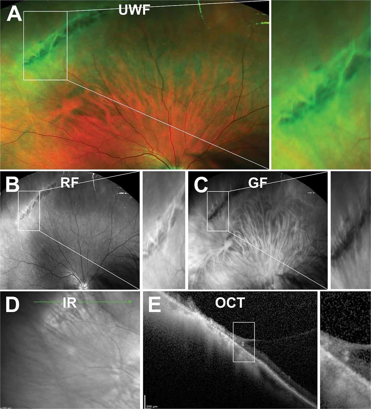
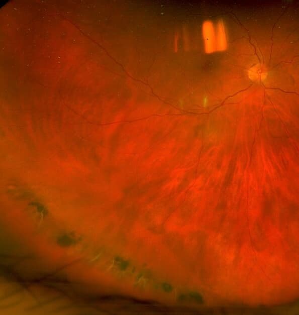
Lesions appear as spindle-shaped or linear areas of retinal thinning, typically oriented circumferentially, with a network of white, sclerosed retinal vessels crossing through them, which is what gives the degeneration its lattice or crosshatched name.
Overlying pigmentation, either hyperpigmented or hypopigmented, is common within and around the lesion. Yellow-white flecks can be seen scattered through the area in some cases.
The vitreous overlying a lattice lesion is often liquefied, while the vitreous immediately at its margins remains abnormally adherent, a combination that is central to why these lesions are a recognised source of retinal breaks.
Pathophysiology
The retina within a lattice lesion is thinned, with loss of the inner retinal layers and, in more advanced lesions, occasionally full-thickness atrophic holes forming within the lesion itself.
Abnormally strong vitreoretinal adhesion at the margins of the lesion is the key mechanical feature. When posterior vitreous detachment occurs later in life, traction is concentrated exactly at these adherent margins rather than being distributed evenly across the retina.
This localised traction at the lesion edge is what can tear the thinned retina, producing a horseshoe-shaped retinal tear at the margin of an otherwise stable-looking lattice lesion. Atrophic holes within the lesion itself, by contrast, are usually not traction-related and carry a comparatively lower risk of progressing to detachment.
Fundus Explorer Pro
Photograph the retinal findings described here with the phone already in your pocket — 22 D optics and built-in illumination in one handheld unit.
From Choroida — the team behind this siteEpidemiology
Lattice degeneration is present in a meaningful proportion of the general population and is more common in myopic eyes, particularly with increasing axial length.
It is bilateral in a substantial proportion of affected patients, which is a practical reason to examine the fellow eye carefully once lattice is found in one.
Lesions are most often located in the superotemporal and inferotemporal quadrants, though they can occur anywhere in the peripheral retina.
Clinical Presentation
Lattice degeneration itself is asymptomatic and is typically found incidentally during a routine dilated peripheral retinal examination, often in a myopic patient or one undergoing screening for another reason.
Symptoms only arise if a complication develops. New flashes and floaters suggest either acute posterior vitreous detachment with traction at a lattice margin, or an actual retinal tear, and warrant prompt dilated examination.
A visual field defect, described as a curtain or shadow, suggests that a retinal detachment has already developed and needs urgent same-day assessment.
Risk Factors for Complications
- Myopia, with higher degrees of axial elongation associated with more extensive lattice degeneration and greater overall retinal detachment risk
- Lattice degeneration in the fellow eye of a patient who has already had a retinal detachment, which substantially raises the risk to that eye
- A family history of retinal detachment
- Ocular trauma, which can precipitate acute vitreous traction at a lattice margin
- Aphakia or pseudophakia, since cataract surgery is associated with an increased rate of posterior vitreous detachment and subsequent retinal breaks
Differential Diagnosis
- Paving-stone (cobblestone) degeneration, discrete pale, well-demarcated areas near the ora serrata, generally considered lower risk and not associated with vitreoretinal traction in the same way
- Snail-track degeneration, a related but distinct pattern of glistening white tracks without the crosshatched vessel pattern of true lattice
- White-with-pressure or white-without-pressure changes, seen on scleral depression and representing a different, generally benign, vitreoretinal interface finding
- A retinal tear without underlying lattice, which can occur in an otherwise normal-appearing peripheral retina and needs to be distinguished from a tear specifically at the edge of a lattice lesion
Evaluation
Dilated indirect ophthalmoscopy with scleral depression is the standard way to examine the full extent of the peripheral retina, including areas that are difficult to visualise without depression.
Every lattice lesion found should be examined carefully for an associated retinal break, since the presence of a break, not simply the presence of lattice, is what changes management from observation to treatment.
Documentation with a retinal drawing or wide-field photography is useful for future comparison, particularly in patients being followed over time or those with high-risk features.
Management
Asymptomatic Lattice Degeneration
Most lattice degeneration without an associated break or symptoms is managed with observation and patient education about the symptoms of a retinal tear or detachment, rather than prophylactic treatment. Routine treatment of every lattice lesion found incidentally is not supported by the evidence and is not standard practice.
Lattice With a Retinal Tear or Symptomatic Presentation
Laser retinopexy or cryotherapy is used to wall off a retinal tear found within or at the margin of lattice degeneration, aiming to create a chorioretinal adhesion that prevents subretinal fluid from tracking through the break.
Selected Higher-Risk Situations
Prophylactic treatment of lattice degeneration without a break is sometimes considered in specific higher-risk circumstances, such as the fellow eye of a patient with a prior retinal detachment, or before intraocular surgery in an eye with extensive lattice, though this remains a matter of individual clinical judgement rather than a universal recommendation.
Patient Counselling
Patients found to have lattice degeneration should understand that the finding itself does not require treatment in most cases, but that they should seek urgent assessment for any new flashes, floaters, or a shadow in their peripheral vision.
This counselling matters more than any specific treatment decision, since early presentation with a symptomatic tear, before it progresses to detachment, is what keeps outcomes good.
Prognosis
The great majority of patients with lattice degeneration never develop a retinal detachment, and observation alone is the right approach for most.
When a retinal tear does develop at a lattice margin and is treated promptly with laser or cryotherapy, the chorioretinal adhesion it creates is generally effective at preventing progression to detachment.
Delayed presentation with an established detachment carries a worse visual prognosis than a tear caught and treated before subretinal fluid accumulates, which is the central argument for patient education over blanket prophylactic treatment of every lesion found.


Document what you see
Two smartphone imaging tools built for everyday clinic use — one for the slit lamp, one for the fundus.
From Choroida — the team behind this siteReferences
- Byer NE. Long-term natural history of lattice degeneration of the retina. Ophthalmology. 1989.
- Wilkinson CP. Interventions for asymptomatic retinal breaks and lattice degeneration for preventing retinal detachment. Cochrane Database of Systematic Reviews. 2014.
- Folk JC, Arrindell EL, Klugman MR. The fellow eye of patients with phakic lattice retinal detachment. Ophthalmology. 1989.
- Lattice Degeneration. EyeWiki, American Academy of Ophthalmology.
- Lattice Degeneration of the Retina. StatPearls, NCBI Bookshelf.
Test yourself
A few questions straight from this article.
-
Where in the retina is lattice degeneration most commonly located?
Lattice degeneration is a peripheral retinal thinning with criss-crossing sclerosed vessels over atrophic retina, sited most commonly between the equator and the ora serrata. -
How does the vitreous typically behave over and around a lattice degeneration lesion?
The vitreous overlying lattice is often liquefied while remaining abnormally adherent at the lesion margins, and this combination is why lattice is a recognised source of retinal breaks. -
Which retinal break typically results from posterior vitreous detachment in an eye with lattice degeneration?
When posterior vitreous detachment occurs, traction concentrates at the abnormally adherent lesion margins rather than spreading evenly, tearing the thinned retina and producing a horseshoe tear at the edge. -
How do atrophic holes forming within a lattice lesion differ from tears at its margin?
Atrophic holes within the lesion are generally not traction-related and carry a comparatively lower risk of progressing to retinal detachment than a traction tear at the margin. -
Which finding changes management of lattice degeneration from observation to treatment?
Every lattice lesion is examined for an associated break, because it is the presence of a break, not lattice itself, that converts management from observation to treatment. -
Which peripheral finding is distinguished from lattice by glistening white tracks without a crosshatched vessel pattern?
Snail-track degeneration is a related but distinct pattern of glistening white tracks that lacks the crosshatched sclerosed-vessel network defining true lattice. -
Which examination technique is standard for assessing the full extent of lattice degeneration?
Dilated indirect ophthalmoscopy with scleral depression is the standard method for viewing the full peripheral retina, including areas difficult to visualise without depression. -
How does laser retinopexy protect an eye with a retinal tear at a lattice margin?
Laser retinopexy or cryotherapy walls off the tear by creating a chorioretinal adhesion that stops subretinal fluid tracking through the break. -
In which situation is prophylactic treatment of lattice degeneration without a break sometimes considered?
Prophylaxis without a break is reserved for selected higher-risk circumstances such as the fellow eye after a retinal detachment, or before intraocular surgery in an eye with extensive lattice. -
Which symptom in a patient known to have lattice degeneration indicates urgent same-day assessment?
A visual field defect described as a curtain or shadow suggests a retinal detachment has already developed and needs urgent same-day assessment, whereas new flashes and floaters warrant prompt dilated examination.

Writes and reviews the clinical articles on Educate. Every post is checked against peer-reviewed sources before it goes live.


