
Laser retinopexy is the use of focal laser photocoagulation to seal a retinal break before it can progress to a rhegmatogenous retinal detachment.
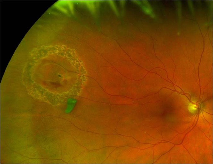
It works by creating a controlled chorioretinal scar around the break, not by treating any subretinal fluid that may already be present.
That distinction shapes almost everything about when laser retinopexy is appropriate and when it is not enough on its own.
Mechanism
The laser burn causes thermal coagulation of the retinal pigment epithelium and adjacent choroid, and as this heals it forms a firm adhesion between the retina and the underlying tissue.
That adhesion typically takes several days to reach full strength, which is why a freshly lasered break is still considered at risk until the chorioretinal scar has matured.
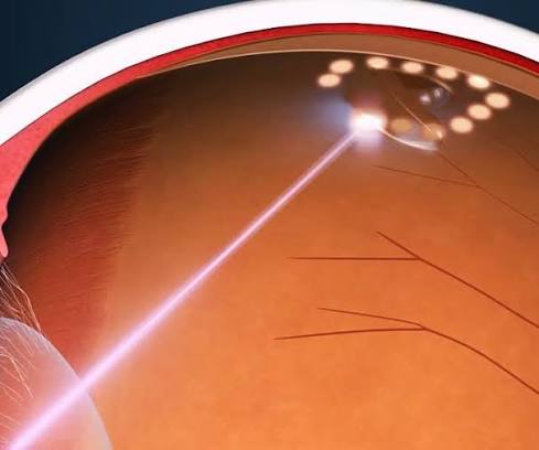
The goal is a ring of confluent burns surrounding the break rather than a single spot, since an incomplete ring leaves a gap through which fluid can still track underneath the retina.
Indications
Laser retinopexy is the treatment of choice for a symptomatic retinal tear with no more than a trace of surrounding subretinal fluid, and for prophylactic treatment of high-risk asymptomatic lesions such as a horseshoe tear found incidentally on a dilated exam.
- Acute symptomatic horseshoe or operculated tears without an associated detachment
- Asymptomatic tears with a high risk profile, including lattice degeneration with an associated break, or a tear in the fellow eye of a patient who has already had a detachment
- Adjunctive treatment around a drainage site or residual break after primary retinal detachment surgery
- Demarcation of a chronic, stable, shallow detachment to wall off further progression when full reattachment surgery is not otherwise indicated
Atrophic round holes without traction, and lesions like white-without-pressure that carry a low intrinsic risk of detachment, are generally observed rather than treated, since the risk of treatment can outweigh the risk of the lesion itself.
Fundus Explorer Pro
Photograph the retinal findings described here with the phone already in your pocket — 22 D optics and built-in illumination in one handheld unit.
From Choroida — the team behind this siteLaser Versus Cryotherapy
Cryotherapy achieves the same biological goal, a chorioretinal adhesion, through freezing applied to the external scleral surface rather than light applied through the pupil.
Laser is generally preferred when the break is visible and accessible through a clear media and a dilated pupil, since it is more precise, better tolerated, and easier to titrate burn by burn.
Cryotherapy remains useful for very peripheral breaks near the ora serrata that are difficult to reach with a laser delivery system, and it is the natural choice when a scleral buckle procedure is already being performed and the sclera is already exposed.
Cryotherapy also tends to cause more breakdown of the blood-retinal barrier than laser does, which some surgeons weigh when a patient already has risk factors for proliferative vitreoretinopathy.
Technique
Treatment is usually performed at the slit lamp using an indirect laser delivery system or a contact lens, with the break centered and a continuous ring of burns placed two to three rows deep around its entire circumference.
Burn intensity is set to produce a moderate, gray-white retinal reaction rather than an intense white burn, since overly aggressive treatment increases the risk of a full-thickness retinal hole forming at the burn site itself.
Multiple sessions are sometimes needed to complete a full barrier, particularly around a large horseshoe tear, and the surgeon confirms a closed ring before considering the treatment finished.
Most patients tolerate the procedure well with only topical anesthesia, though treatment near the horizontal meridian, close to the long posterior ciliary nerves, tends to be more uncomfortable and occasionally needs a peribulbar or sub-Tenon’s injection.
What Laser Retinopexy Cannot Do
Laser retinopexy does not reattach retina that has already separated from the pigment epithelium, because the burn cannot create an adhesion across a gap filled with subretinal fluid.
A break with more than a minimal cuff of surrounding fluid needs a reattachment procedure, whether scleral buckle, vitrectomy, or pneumatic retinopexy, and laser alone in that setting simply fails to hold.
This is the most common pitfall in managing peripheral retinal breaks: treating a break that already has a meaningful rim of subretinal fluid with laser alone, only to have the detachment progress despite an apparently complete-looking burn pattern.
Follow-Up and Failure
Patients are typically re-examined within one to two weeks to confirm the burns have matured into visible pigmented scars and that no new fluid has accumulated.
New symptoms after treatment, particularly increasing flashes, a new field defect, or a shadow encroaching from the periphery, warrant prompt re-examination rather than waiting for the scheduled follow-up.
Treatment failure is more likely when the initial ring was incomplete, when a second unrecognized break was present elsewhere, or when vitreous traction at the site continues to pull on the retina despite an adequate chorioretinal scar.
A dilated fundus exam of the fellow eye is also worth repeating at follow-up, since a patient who develops one symptomatic tear has an elevated lifetime risk of a break in the other eye as well.


Document what you see
Two smartphone imaging tools built for everyday clinic use — one for the slit lamp, one for the fundus.
From Choroida — the team behind this siteReferences
- Wilkinson CP, Rice TA. Michels’ Retinal Detachment.
- American Academy of Ophthalmology. Basic and Clinical Science Course, Section 12: Retina and Vitreous.
- Wilkinson CP. Evidence-based analysis of prophylactic treatment of asymptomatic retinal breaks and lattice degeneration. Ophthalmology.
- Byer NE. Long-term natural history of lattice degeneration of the retina. Ophthalmology.
Test yourself
A few questions straight from this article.
-
What does laser retinopexy actually achieve around a peripheral retinal break?
Laser retinopexy seals a break by walling it off with a chorioretinal scar. It does nothing to subretinal fluid that is already present, which is why fluid volume decides whether laser alone is enough. -
Which tissue change produces the retinal adhesion created by laser retinopexy?
The laser burn thermally coagulates the retinal pigment epithelium and the choroid beneath it, and the healing response binds the retina firmly to the underlying tissue. -
How soon does the chorioretinal adhesion after laser retinopexy reach full strength?
The adhesion needs several days to mature, so a freshly lasered break is still regarded as at risk until the chorioretinal scar has fully formed. -
What burn pattern should surround a retinal tear treated with laser retinopexy?
A confluent ring two to three rows deep must encircle the whole break. Any gap in the ring leaves a channel through which fluid can still track under the retina. -
Laser retinopexy alone suits a symptomatic retinal tear with how much surrounding subretinal fluid?
Laser retinopexy is the treatment of choice for a symptomatic tear with at most a trace of surrounding subretinal fluid. Anything more needs a reattachment procedure. -
A peripheral break with more than a minimal cuff of subretinal fluid requires which management?
A burn cannot bridge a gap filled with subretinal fluid, so laser simply fails to hold. Scleral buckle, vitrectomy or pneumatic retinopexy is needed instead. -
In which situation is cryotherapy preferred over laser for sealing a retinal break?
Cryotherapy reaches breaks too anterior for a laser delivery system, and it is the natural choice when a scleral buckle is already being placed and the sclera is exposed. -
Which peripheral lesion is generally observed rather than sealed with laser retinopexy?
Atrophic round holes without traction, like white-without-pressure, carry a low intrinsic detachment risk, so the hazard of treating them can outweigh the hazard of leaving them. -
What complication follows excessively intense laser burns during retinopexy for a retinal tear?
Burns are titrated to a moderate grey-white reaction rather than an intense white one, because overly aggressive treatment can itself create a full-thickness hole at the treated spot. -
Which finding at the one- to two-week visit confirms successful laser retinopexy of a tear?
Re-examination at one to two weeks looks for burns that have matured into pigmented scars and confirms that no new subretinal fluid has appeared.
