
Ultra-widefield retinal imaging captures a much larger portion of the fundus in a single image than conventional fundus cameras, which usually record 30 to 50 degrees.
Scanning laser systems can image up to about 200 degrees, roughly 80 percent of the retinal surface, in one capture.
This changes what can be documented in diabetic retinopathy, vascular occlusion, uveitis, and peripheral degenerations, although the extra field brings its own limitations.
What Is Ultra-Widefield Retinal Imaging?
Conventional fundus photography records the posterior pole and part of the midperiphery.
Widefield systems extend this coverage, and ultra-widefield systems reach beyond the vortex vein ampullae toward the ora serrata.
The International Widefield Imaging Study Group proposed definitions based on anatomic landmarks and not on degrees, since angular measurements vary with axial length and projection.
Main technologies include:
- Scanning laser ophthalmoscopy (such as Optos), with an ellipsoid mirror that allows capture of a large field through an undilated pupil
- Widefield fundus cameras that capture 100 to 133 degrees in a single image and extend further through montage
- Contact and noncontact widefield systems used in operating rooms and neonatal units
- Portable and smartphone-based devices, which offer smaller fields but greater accessibility
Pseudocolor images combine red and green laser channels.
The green channel images the sensory retina, and the red channel penetrates deeper into the RPE and choroid.
Clinical Uses
Diabetic Retinopathy
Diabetic lesions such as hemorrhages, microaneurysms, and neovascularization often lie outside the standard seven fields.
In a large study of ultra-widefield imaging, predominantly peripheral lesions were found in a meaningful minority of eyes and were associated with a higher risk of progression over several years.
Including the periphery changed the retinopathy severity level in some eyes, which can alter follow-up intervals (see leaky vessels on angiography).
Widefield angiography also identifies areas of capillary nonperfusion, and eyes with more nonperfusion have a higher risk of progression.
Retinal Vein and Artery Occlusion
Widefield fluorescein angiography shows the extent of ischemia and the location of neovascularization, which helps plan laser and injections (see central retinal vein occlusion).
Uveitis and Vasculitis
Peripheral vasculitis, leakage, and snowbanking are seen more clearly, and disease activity can be monitored without scleral depression.
Retinal Detachment and Peripheral Lesions
Ultra-widefield imaging documents retinal breaks, lattice degeneration, and detachments, and it helps track them over time (see lattice degeneration).
Tumors and Pigmented Lesions
Peripheral nevi, melanoma, and congenital lesions can be photographed and monitored for growth.
Pediatric Retina
Contact widefield cameras are used in retinopathy of prematurity, familial exudative vitreoretinopathy, and Coats disease (see Coats disease).
Screening and Telemedicine
Widefield images obtained through an undilated pupil support diabetic screening programs and remote reading, and artificial intelligence tools are being trained on them.
Fundus Explorer Pro
Photograph the retinal findings described here with the phone already in your pocket — 22 D optics and built-in illumination in one handheld unit.
From Choroida — the team behind this siteAdditional Modalities
Ultra-widefield autofluorescence shows RPE health in the periphery, including in inherited disease and in AMD.
Widefield swept-source OCT and montage OCT extend structural imaging to the periphery, and widefield OCT angiography is emerging.
Limitations and Artifacts
- Peripheral distortion: the two-dimensional image of a curved surface stretches the periphery, so distances and areas are not accurate unless corrected
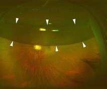
- Eyelid and eyelash artifacts: the superior and inferior fundus are often obscured
- Pseudocolor rendering: colors differ from real fundus colors, and some lesions look different from what an examiner sees
- Limited stereopsis: depth cues are absent
- Limited view of the far periphery: the anterior periphery near the ora serrata may be inadequately imaged, and it does not replace scleral depression
- Media opacity: cataract and vitreous haze reduce image quality
- Cost and access: systems are expensive, and images require experienced graders
An ultra-widefield image is a document and a screening aid, and it does not replace a dilated examination in patients with symptoms of retinal tears or detachment.
Photography or Angiography?
Color or pseudocolor widefield photography suits documentation and follow-up of visible lesions such as hemorrhages, tumors, and detachments.
Autofluorescence adds information about RPE health without dye.
Widefield fluorescein angiography is chosen when perfusion and leakage matter, for example in vein occlusion, diabetic retinopathy with suspected peripheral neovascularization, and vasculitis.
The choice depends on whether the answer will change treatment, since angiography takes longer and carries the small risks of the dye.
Practical Tips
- Ask the patient to look in the requested directions to capture the periphery, and repeat the capture when lashes obstruct the field
- Use widefield fluorescein angiography when nonperfusion or peripheral leakage will change management
- Compare images taken on the same device over time, since device differences make comparison unreliable
- Correct for projection when measuring lesions, or use software designed for the purpose
- Record the device and settings in the chart
Future Directions
Automated grading for diabetic retinopathy, quantification of nonperfusion, and machine-learning detection of peripheral lesions are active areas of research.
Standardized terminology will help compare studies and translate results into practice.


Document what you see
Two smartphone imaging tools built for everyday clinic use — one for the slit lamp, one for the fundus.
From Choroida — the team behind this siteReferences
- Nagiel A, Lalane RA, Sadda SR, Schwartz SD. Ultra-widefield fundus imaging: a review of clinical applications and future trends. Retina. 2016;36:660-678.
- Choudhry N, Duker JS, Freund KB, et al. Classification and guidelines for widefield imaging: recommendations from the International Widefield Imaging Study Group. Ophthalmol Retina. 2019;3:843-849.
- Silva PS, Cavallerano JD, Haddad NM, et al. Peripheral lesions identified on ultrawide field imaging predict increased risk of diabetic retinopathy progression over 4 years. Ophthalmology. 2015;122:949-956.
- Silva PS, Dela Cruz AJ, Ledesma MG, et al. Diabetic retinopathy severity and peripheral lesions are associated with nonperfusion on ultrawide field angiography. Ophthalmology. 2015;122:2465-2472.
- Patel SN, Shi A, Wibbelsman TD, Klufas MA. Ultra-widefield retinal imaging: an update on recent advances. Ther Adv Ophthalmol. 2020;12:2515841419899495.
Test yourself
A few questions straight from this article.
-
How much of the retina can a scanning laser ultra-widefield system capture in one image?
Scanning laser systems can image up to about 200 degrees, roughly 80 percent of the retinal surface, in a single capture. Conventional fundus cameras usually record only 30 to 50 degrees. -
Why did the International Widefield Imaging Study Group define widefield imaging by anatomic landmarks rather than degrees?
The group based its definitions on anatomic landmarks rather than degrees, because angular measurements vary with axial length and projection. Ultra-widefield systems reach beyond the vortex vein ampullae toward the ora serrata. -
In pseudocolor ultra-widefield images, what does the red laser channel display?
Pseudocolor images combine red and green laser channels. The green channel images the sensory retina while the red channel penetrates deeper into the RPE and choroid. -
What feature lets scanning laser ophthalmoscopy image a large field through an undilated pupil?
Scanning laser ophthalmoscopy uses an ellipsoid mirror that allows capture of a large field through an undilated pupil, which is why it suits diabetic screening and remote reading programmes. -
What effect can including the retinal periphery have on diabetic retinopathy assessment?
Diabetic lesions often lie outside the standard seven fields, and including the periphery changed the retinopathy severity level in some eyes, which can alter follow-up intervals. Predominantly peripheral lesions carried a higher risk of progression. -
What does widefield fluorescein angiography add in diabetic retinopathy?
Widefield angiography identifies areas of capillary nonperfusion, and eyes with more nonperfusion carry a higher risk of progression. Autofluorescence is what adds information on peripheral RPE health without dye. -
Why are distances and areas unreliable on an uncorrected ultra-widefield image?
Rendering a curved retinal surface as a two-dimensional image stretches the periphery, so distances and areas are not accurate unless corrected. Measurement needs projection correction or dedicated software. -
Which part of the fundus is most often obscured by artifact on ultra-widefield imaging?
Eyelid and eyelash artifacts often obscure the superior and inferior fundus. Repeating the capture, and asking the patient to look in the requested directions, helps recover the obstructed field. -
What is the role of ultra-widefield imaging in a patient with symptoms of a retinal tear?
An ultra-widefield image is a document and a screening aid. The anterior periphery near the ora serrata may be inadequately imaged, so it does not replace scleral depression or a dilated examination in symptomatic patients. -
Why should serial ultra-widefield images be compared on the same device?
Images should be compared on the same device over time, since device differences make comparison unreliable. The device and settings should also be recorded in the chart.

Writes and reviews the clinical articles on Educate. Every post is checked against peer-reviewed sources before it goes live.


