
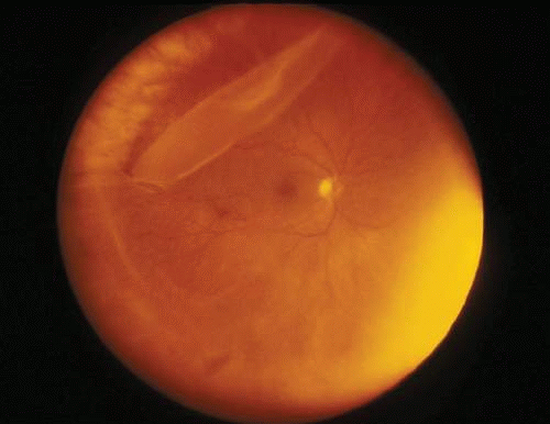
A giant retinal tear is defined as a full-thickness retinal break extending 90 degrees or more of the circumference of the eye.
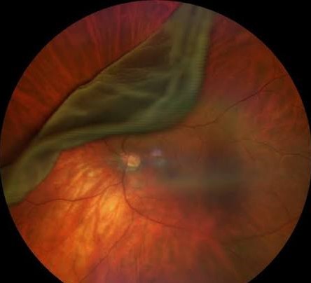
It represents one of the most surgically challenging forms of rhegmatogenous retinal detachment, both because of the sheer size of the break and because the detached posterior flap of retina characteristically rolls and folds on itself, making it a different technical problem from managing a standard, small horseshoe tear.
Why Giant Tears Behave Differently
Unlike a small retinal break, where the surrounding retina remains stable and tacked in place, the posterior edge of a giant tear is entirely free of any anterior attachment along its full extent, allowing it to fold over on itself (the retina “flipping” posteriorly) under the influence of gravity and vitreous traction.
This flap mobility is the central technical challenge of giant tear surgery, because flattening and reattaching the retina requires unfolding and repositioning a large, mobile piece of tissue rather than just sealing a discrete, stable break, and it is why the surgical techniques and instrumentation used differ meaningfully from those needed for a standard, small retinal tear.
Causes and Risk Factors
- Blunt ocular trauma — a major cause, particularly in younger patients, from the sudden anteroposterior compression and equatorial expansion of the globe that trauma produces
- High myopia, with its associated peripheral retinal thinning and weaker vitreoretinal adhesion in some areas
- Stickler syndrome and other inherited vitreoretinopathies, which carry a substantially elevated risk of giant retinal tear, sometimes bilaterally and at a young age, and are discussed in more detail in this site’s dedicated coverage of Stickler syndrome
- Prior giant retinal tear in the fellow eye — a recognized and important risk factor for fellow-eye involvement, given the shared underlying predisposing anatomy or genetic condition, warranting closer surveillance and sometimes prophylactic treatment of the fellow eye
- Spontaneous occurrence without an identifiable predisposing factor, in a minority of cases
Fundus Explorer Pro
Photograph the retinal findings described here with the phone already in your pocket — 22 D optics and built-in illumination in one handheld unit.
From Choroida — the team behind this siteClinical Presentation
Patients present with the sudden onset of flashes, a shower of floaters, and a rapidly progressive visual field defect or curtain, often more dramatic and more rapidly progressive than the presentation of a typical, smaller retinal detachment, reflecting how quickly a giant tear can lead to extensive retinal detachment given the size of the break.
Vision loss can be profound if the detachment has already progressed to involve the macula by the time of presentation, which, given the rapid progression typical of giant tears, is not uncommon.
Exam Findings
- A large, circumferential retinal break, often with the posterior flap visibly folded or rolled over itself, sometimes obscuring clear visualization of the full extent of the tear on initial exam
- Extensive associated retinal detachment, frequently already involving or threatening the macula given how quickly giant tears tend to progress
- Vitreous hemorrhage in a subset of cases, particularly when trauma is the underlying cause, which can further complicate visualization and surgical planning
- Signs of an underlying predisposing condition when relevant — high myopia, or the systemic features of Stickler syndrome (distinctive facial features, joint hypermobility, hearing loss) in appropriate cases
Differential Diagnosis
- Standard rhegmatogenous retinal detachment from a smaller horseshoe tear — a discrete, stable break rather than an extensive, circumferential, mobile-flap tear
- Retinal dialysis — a full-thickness break at the ora serrata, typically from trauma, but anatomically and surgically distinct from a giant tear located more posteriorly
- Tractional retinal detachment — a fundamentally different mechanism (membrane contraction rather than a break), discussed in its own dedicated article on this site, with a different fundus appearance (concave, immobile) rather than the convex, mobile detachment typical of a giant tear
Surgical Management
Pars plana vitrectomy is the standard approach, with particular surgical attention to thoroughly removing vitreous traction on the mobile posterior flap, often facilitated by perfluorocarbon liquid.
This is used specifically in giant tear surgery to “unfold” and flatten the rolled posterior flap and stabilize the retina intraoperatively in a way that greatly simplifies an otherwise technically difficult maneuver.
Endolaser is applied along the tear margins once the retina is flattened.
Long-acting gas or silicone oil tamponade is used postoperatively, with the choice between them influenced by tear size, patient factors, and the surgeon’s assessment of how reliably the patient can maintain required postoperative positioning.
Given the elevated risk of fellow-eye involvement in patients with an underlying predisposing condition such as Stickler syndrome, prophylactic examination — and sometimes prophylactic treatment of high-risk areas — of the fellow eye is an important part of comprehensive management, extending the scope of care beyond just the initially affected eye.
Prognosis
Anatomic success rates for giant retinal tear repair have improved substantially with modern vitrectomy techniques and perfluorocarbon liquid use, though outcomes remain more variable than for standard retinal detachment repair.
Final visual acuity depends heavily on whether the macula was involved before surgery and for how long.
Proliferative vitreoretinopathy, a scarring response that can cause recurrent detachment after apparently successful initial repair, is a particular concern in giant tear cases given the extent of retinal manipulation often required during surgery.
Postoperative monitoring specifically watches for this complication, since early detection of recurrent traction or detachment allows for prompter intervention than waiting for a scheduled routine follow-up visit to reveal a problem that has already progressed significantly.


Document what you see
Two smartphone imaging tools built for everyday clinic use — one for the slit lamp, one for the fundus.
From Choroida — the team behind this siteReferences
- Freeman HM. Fellow eyes of giant retinal breaks. Transactions of the American Ophthalmological Society.
- Ang GS, Townend J, Lois N. Epidemiology of giant retinal tears in the United Kingdom. Investigative Ophthalmology & Visual Science.
- American Academy of Ophthalmology. Basic and Clinical Science Course, Section 12: Retina and Vitreous.
Test yourself
A few questions straight from this article.
-
A giant retinal tear is defined by a full-thickness break extending at least how far?
A giant retinal tear is a full-thickness retinal break extending 90 degrees or more of the circumference of the eye. -
What makes a giant retinal tear behave differently from a small horseshoe tear?
The posterior edge of a giant tear has no anterior attachment along its full extent, so it folds over on itself under gravity and vitreous traction. That flap mobility is the central technical challenge of surgery. -
How does blunt ocular trauma produce a giant retinal tear?
Blunt trauma is a major cause, particularly in younger patients, through the sudden anteroposterior compression and equatorial expansion of the globe that the impact produces. -
Which inherited condition carries a substantially raised risk of giant retinal tear?
Stickler syndrome and other inherited vitreoretinopathies carry a substantially elevated risk of giant retinal tear, sometimes bilaterally and at a young age. -
Why does a patient with a giant retinal tear need close surveillance of the fellow eye?
A prior giant retinal tear in one eye is a recognized risk factor for fellow-eye involvement, given the shared predisposing anatomy or genetic condition, warranting closer surveillance and sometimes prophylactic treatment. -
How does a giant retinal tear typically present?
Patients present with sudden flashes, a shower of floaters, and a rapidly progressive field defect or curtain, often more dramatic than a typical smaller detachment given how quickly a giant tear progresses. -
How does a tractional retinal detachment differ in appearance from a giant tear detachment?
Tractional detachment arises from membrane contraction rather than a break, and has a concave, immobile appearance rather than the convex, mobile detachment typical of a giant tear. -
What is perfluorocarbon liquid used for during giant retinal tear surgery?
Perfluorocarbon liquid unfolds and flattens the rolled posterior flap and stabilizes the retina intraoperatively, after which endolaser is applied along the tear margins and gas or silicone oil provides tamponade. -
Which complication is a particular concern after giant retinal tear repair?
Proliferative vitreoretinopathy, a scarring response causing recurrent detachment after apparently successful repair, is a particular concern given the extent of retinal manipulation often required, so postoperative monitoring watches for it specifically. -
What most strongly determines final visual acuity after giant retinal tear repair?
Final visual acuity depends heavily on whether the macula was involved before surgery and for how long, even though anatomic success rates have improved substantially with modern vitrectomy and perfluorocarbon liquid.

Writes and reviews the clinical articles on Educate. Every post is checked against peer-reviewed sources before it goes live.


