
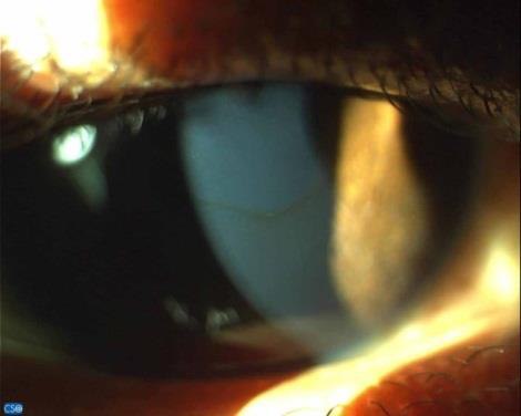
The hardest keratoconus to catch is the one that still sees 6/6.
By the time a cornea shows a visible cone, the diagnosis is easy. The clinical challenge — and the reason it is so often missed for years — is the early eye: a young patient whose glasses never quite work, whose astigmatism keeps shifting, and whose examination looks almost normal.
Keratoconus is the most common corneal ectasia, and recognizing it early now changes the outcome, because cross-linking can halt progression before vision is lost.
What Is Keratoconus?
Keratoconus is a progressive, non-inflammatory thinning and steepening of the cornea, which bulges forward into a cone shape. The ectasia is usually bilateral but asymmetric, and it typically involves the inferior or central cornea.
The distorted cornea generates irregular astigmatism and higher-order aberrations that spectacles cannot fully correct. That single fact — vision limited by an irregular surface rather than a simple refractive error — explains most of the disease’s clinical behavior.
Epidemiology and Associations
Keratoconus usually begins around puberty and progresses through the second and third decades before tending to stabilize later in adulthood. Onset in a teenager generally signals a more aggressive course.
- Eye rubbing is the most important modifiable risk factor and is strongly associated with progression.
- Atopy — allergic eye disease, eczema, and asthma — is common, partly through chronic rubbing.
- Systemic associations include Down syndrome and connective-tissue disorders.
- A positive family history is present in a substantial minority.
Asking specifically about eye rubbing is not a formality — it is one of the few levers that can slow the disease.
All-fit Slit-Lamp Adapter
Record and share exactly what you see at the slit lamp. One adapter fits any slit lamp or surgical microscope — and any smartphone.
From Choroida — the team behind this siteClinical Presentation
The story is often more revealing than the early examination.
- Progressive blurring and distortion not fully corrected by glasses.
- Frequent changes of spectacle prescription, especially rising and rotating astigmatism.
- Monocular ghosting, glare, and haloes, worse at night.
- A history of vigorous eye rubbing, often with atopy.
When a young patient’s astigmatism keeps shifting and best-corrected vision quietly falls, keratoconus should be actively excluded rather than assumed absent.
Slit-Lamp and Clinical Signs
Classic signs appear as the disease advances, and knowing them helps confirm a suspicion raised by topography.
- Stromal thinning at the apex of the cone.
- Fleischer ring — an iron deposit encircling the base of the cone.
- Vogt striae — fine vertical stress lines in the deep stroma that fade with gentle pressure on the globe.
- Munson sign — a V-shaped bulge of the lower lid on downgaze in advanced cones.
- Corneal hydrops — sudden painful edema from a break in Descemet membrane, with acute clouding of vision.
These signs are useful when present, but their absence never rules out early keratoconus — that is the job of imaging.
Diagnostic Evaluation
Modern diagnosis is driven by corneal imaging rather than the slit lamp alone.
- Corneal topography shows inferior steepening and an asymmetric bowtie — often the earliest objective clue.
- Tomography (e.g., Scheimpflug imaging) adds posterior-surface elevation and a full pachymetry map, and detects subclinical disease topography can miss.
- Retinoscopy may reveal a scissoring reflex; keratometry shows steep, irregular readings.
- Serial scans are essential, because documented progression is what justifies treatment.
Screening the fellow eye and any refractive-surgery candidate for subclinical ectasia is now a standard part of the workup.
Differential Diagnosis
- Pellucid marginal degeneration — inferior thinning with a “crab-claw” topography and against-the-rule astigmatism.
- Keratoglobus — diffuse, global thinning rather than a focal cone.
- Post-refractive-surgery ectasia — a similar picture after LASIK.
- Contact-lens-induced warpage — reversible distortion that resolves after lens removal.
Separating these matters, because their management and prognosis diverge sharply from classic keratoconus.
Management
Treatment now has two distinct goals: stop progression and rehabilitate vision.
- Stop eye rubbing and treat any underlying atopy — the simplest and most overlooked intervention.
- Corneal collagen cross-linking (CXL) stiffens the cornea and is the key treatment to halt documented progression, ideally before significant vision loss.
- Spectacles or soft lenses suffice in mild disease.
- Rigid gas-permeable, hybrid, or scleral contact lenses correct the irregular surface as vision worsens and are the mainstay of visual rehabilitation.
- Intracorneal ring segments can reshape selected corneas.
- Corneal transplantation (deep anterior lamellar or penetrating keratoplasty) is reserved for advanced scarring or intolerance to lenses.
The central shift in modern care is timing: cross-linking early preserves the cornea, while lenses and surgery mainly restore vision already compromised.
Prognosis
Most patients retain functional vision with contact lenses, and only a minority ultimately need a corneal graft. The prognosis has improved substantially where cross-linking is available and disease is caught while still mild.
The determining factor is how early it is found — which makes screening young patients with shifting astigmatism the single most valuable habit.
Would you like to document corneal signs like keratoconus with your smartphone?


Document what you see
Two smartphone imaging tools built for everyday clinic use — one for the slit lamp, one for the fundus.
From Choroida — the team behind this siteReferences
- Santodomingo-Rubido J, Carracedo G, Suzaki A, et al. Keratoconus: an updated review. Contact Lens and Anterior Eye. 2022;45(3):101559.
- Gomes JAP, Tan D, Rapuano CJ, et al. Global consensus on keratoconus and ectatic diseases. Cornea. 2015;34(4):359-369.
- Wollensak G, Spoerl E, Seiler T. Riboflavin/ultraviolet-A-induced collagen crosslinking for the treatment of keratoconus. American Journal of Ophthalmology. 2003;135(5):620-627.
- Davidson AE, Hayes S, Hardcastle AJ, Tuft SJ. The pathogenesis of keratoconus. Eye. 2014;28(2):189-195.
Test yourself
A few questions straight from this article.
-
Which description best fits keratoconus?
Keratoconus is progressive, non-inflammatory thinning and steepening of the cornea that bulges into a cone; it is usually bilateral but asymmetric and typically involves the inferior or central cornea. -
At what stage of life does keratoconus typically begin?
Keratoconus usually starts around puberty and progresses through the second and third decades before tending to stabilise; onset in a teenager generally signals a more aggressive course. -
Which is the most important modifiable risk factor for keratoconus progression?
Eye rubbing is the most important modifiable risk factor and is strongly associated with progression, which is why atopy and rubbing must be asked about and treated directly. -
Why do spectacles fail to fully correct vision in keratoconus?
The distorted cornea produces irregular astigmatism and higher-order aberrations that spectacles cannot fully correct, so vision is limited by surface irregularity rather than simple refractive error. -
The Fleischer ring seen in keratoconus is which finding?
The Fleischer ring is an iron deposit that encircles the base of the cone, one of the classic slit-lamp signs that appear as keratoconus advances. -
Acute corneal hydrops in keratoconus results from which event?
A break in Descemet membrane allows sudden stromal oedema, producing painful corneal swelling with acute clouding of vision. -
Which corneal topography pattern is often the earliest objective clue to keratoconus?
Topography showing inferior steepening and an asymmetric bowtie is often the earliest objective clue; tomography adds posterior elevation and pachymetry and can detect subclinical disease topography misses. -
Which feature distinguishes pellucid marginal degeneration from keratoconus?
Pellucid marginal degeneration shows inferior thinning with a crab-claw topographic pattern and against-the-rule astigmatism, unlike the focal cone of keratoconus. -
Which treatment is used to halt documented progression of keratoconus?
Cross-linking stiffens the cornea and is the key treatment to stop documented progression, ideally before vision is lost; lenses and grafts mainly rehabilitate vision already compromised. -
What is the visual outcome for most patients with keratoconus?
Most patients keep functional vision with contact lenses and only a minority ultimately need a corneal graft, with outcomes better where cross-linking is available and disease is caught while mild.

Writes and reviews the clinical articles on Educate. Every post is checked against peer-reviewed sources before it goes live.


