
Abstract
A corneal ulcer is a disruption or open sore in the outer surface (epithelial) layer of the cornea. Corneal ulcers are frequently caused by some form of eye injury.

Typical healing time for most superficial ulcers is 3-5 days. Ulcers that do not heal within a few days can become infected, and /or extend into the deeper layers of the cornea (called stroma).
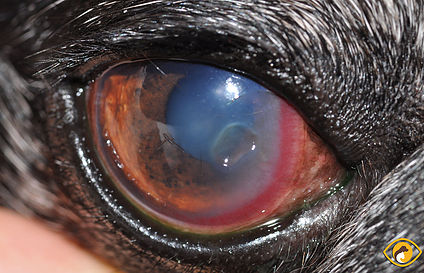
Stromal ulcers can develop for a variety of reasons. Persistent trauma to the surface of the eye by an abnormal eyelash or retained foreign body will delay healing indefinitely.
Conditions such as dry eye, glaucoma, and uveitis can also lead to serious corneal ulcers. Most commonly, ulcers worsen due to infection.
A low population of bacteria is naturally present on the surface of the eye and eyelids. If there is an ulcer on the cornea, these bacteria take the opportunity to colonize (grow) the defect, which can lead to rapid corneal degradation.
Ulcers are referred to as “melting” if the cornea becomes very soft due to enzymatic degradation and breakdown of the corneal collagen. This can lead to rupture of the eye within hours to days of infection and potential loss of the eye.
Stromal Corneal Ulcers Diagnosis
Slit lamp biomicroscopy is used to examine the cornea under a high degree of magnification, and assess the extent and depth of the ulcer.
The slit lamp is also used to look for the potential cause of the ulcer, such as abnormal eyelashes or retained foreign bodies. Schirmer tear testing is also performed to determine if tear production is normal.
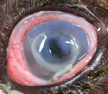
Fluorescein staining is an essential diagnostic test and will bind to exposed corneal stroma, to reveal the size and depth of the ulceration.
Depending on the depth, clinical appearance, and history of the ulceration, additional diagnostic testing such as corneal cytology and corneal culture may be recommended.
Stromal Corneal Ulcers Treatment
Medical management of stromal ulcers involves the frequent instillation of topical antibiotics, as well as a blood product called serum or plasma.
Frequent lubrication with sodium hyaluronate can also aid healing. Additional topical and systemic medications are used to reduce associated eye inflammation and pain.
Our patients are monitored closely and frequently to monitor for progress in healing, as significant changes can occur in a matter of hours.
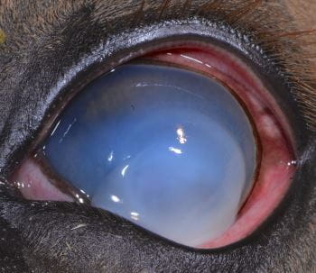
If an ulcer involves 50% or more of the corneal thickness, surgery may be recommended to avoid corneal perforation and loss of vision.
Surgical techniques include the use of corneal grafts or transplants, conjunctival pedicle or island grafts, and other biological graft material to stabilize the cornea. These grafts become incorporated into the patient’s cornea.
Stromal Corneal Ulcers Prognosis
When treatment is sought early, the vast majority of ulcers will heal and at least partial vision will be preserved. These reparative corneal surgeries typically carry a 90% success rate.
As with any surgical procedure, there are potential complications. These include wound dehiscence, graft rejections, persistent infection at the surgical site or inside the eye, corneal scarring, corneal vascularization, corneal mineralization, chronic uveitis, glaucoma, retinal detachment, hyphema (bleeding inside the eye), blindness and loss of the eye.
Our primary goals with medical or surgical treatment of deep corneal ulcers are to preserve the best vision possible in our patients and to control any pain or discomfort they may experience.
HOW TO TAKE SLIT-LAMP EXAM IMAGES WITH A SMARTPHONE?
Smartphone slit-lamp photography is the new advancement in the field of science and technology in which photographs of the desired slit-lamp finding can be taken with smartphones by using the slit-lamp adapters.
