Bleb-associated endophthalmitis is a late-onset intraocular infection that follows filtering glaucoma surgery, and it differs from typical postoperative endophthalmitis in that the risk never fully goes away, sometimes appearing years after the original trabeculectomy.
The thin, avascular bleb created by antimetabolite use is both what makes the surgery work and what leaves a lasting portal for bacteria to enter the eye.
Why the Risk Persists
A functioning filtering bleb is thin-walled and often relatively avascular, especially after mitomycin C or 5-fluorouracil use, and this same thinness that allows good filtration also makes the bleb more permeable to surface organisms.
Unlike routine postoperative endophthalmitis, which clusters in the days after surgery, bleb-associated infection can occur at any point in the life of the bleb, and the annual risk, while low for any given year, accumulates over a patient’s lifetime.
Risk Factors
- Inferior or nasal bleb location, which is more exposed to tear film debris than a superior bleb
- Bleb leak or a thin, cystic, avascular bleb appearance
- Antimetabolite use during the original surgery, particularly mitomycin C
- Blepharitis, conjunctivitis, or chronic ocular surface disease
- Contact lens wear over or near the bleb
- Younger age at the time of surgery, since a longer lifetime of bleb exposure raises cumulative risk
All-fit Slit-Lamp Adapter
Record and share exactly what you see at the slit lamp. One adapter fits any slit lamp or surgical microscope — and any smartphone.
From Choroida — the team behind this siteBlebitis Versus Bleb-Associated Endophthalmitis
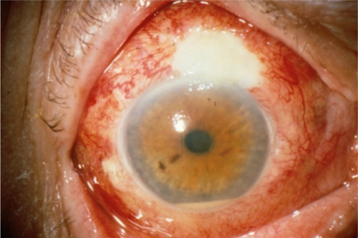
Infection can be confined to the bleb itself, known as blebitis, or can spread into the vitreous cavity as bleb-associated endophthalmitis.
- Blebitis presents with a white, opaque bleb, mild anterior chamber reaction, and preserved vision
- Bleb-associated endophthalmitis adds vitreous involvement, marked pain, significant vision loss, and often a hypopyon
- The organisms responsible tend to be more virulent than in typical postoperative endophthalmitis, with Streptococcus and Haemophilus species reported more often, which explains the frequently aggressive course
Because blebitis can progress rapidly to full endophthalmitis, any patient with a red, painful eye and a history of trabeculectomy needs urgent assessment rather than a routine appointment.
Diagnosis
Diagnosis is clinical, based on the appearance of the bleb, anterior chamber, and vitreous.
- Slit-lamp examination of the bleb for a leak, using a Seidel test with fluorescein
- Assessment of the vitreous for haze, which distinguishes blebitis from endophthalmitis
- Vitreous and aqueous sampling for culture when endophthalmitis is suspected, following the same principles used for other forms of postoperative endophthalmitis
- B-scan ultrasonography when the view is obscured
Management
Blebitis Without Vitreous Involvement
Intensive topical antibiotics, often fortified, along with topical corticosteroids once infection is controlled, are usually sufficient when the vitreous is clear.
Close follow-up is essential, since the situation can worsen quickly.
Bleb-Associated Endophthalmitis
Vitreous involvement is treated as an emergency with intravitreal antibiotics, following the same principles established for postoperative endophthalmitis, and vitrectomy is considered for severe cases with poor vision at presentation.
Systemic antibiotics are sometimes added given the more virulent organism profile typically seen with bleb infections.
After the Acute Episode
Bleb leaks and thin, at-risk blebs may need surgical revision once the infection is controlled, to reduce the risk of recurrence.
Patients should be counseled about ongoing risk and the importance of prompt reporting of any new redness, discharge, or pain around the bleb, for the rest of the bleb’s functional life.
Prevention
- Treating blepharitis and ocular surface disease proactively in patients with a filtering bleb
- Avoiding contact lens wear that overlies the bleb
- Prompt repair of bleb leaks
- Patient education on warning signs, since early presentation improves outcomes substantially
Prognosis
Outcomes are worse than for typical postoperative endophthalmitis, reflecting both the more virulent organisms involved and the sometimes delayed presentation when symptoms are mistaken for minor irritation.
Prompt recognition and treatment give the best chance of preserving vision, and it is why patients with any filtering bleb are told, indefinitely, to treat a new red painful eye as an emergency.


Document what you see
Two smartphone imaging tools built for everyday clinic use — one for the slit lamp, one for the fundus.
From Choroida — the team behind this siteReferences
- Busbee BG. Bleb-associated endophthalmitis: clinical characteristics and visual outcomes. Curr Opin Ophthalmol. 2005;16:170-174.
- Waheed S, Ritterband DC, Greenfield DS, Liebmann JM, Seedor JA, Ritch R. Bleb-related ocular infection in children after trabeculectomy with mitomycin C. Ophthalmology. 1997;104:2117-2120.
- Yamamoto T, Kuwayama Y; Collaborative Bleb-related Infection Incidence and Treatment Study Group. Interim clinical outcomes in the collaborative bleb-related infection incidence and treatment study. Ophthalmology. 2011;118:453-458.
- Kangas TA, Greenfield DS, Flynn HW Jr, Parrish RK 2nd, Palmberg P. Delayed-onset endophthalmitis associated with conjunctival filtering blebs. Ophthalmology. 1997;104:746-752.
Test yourself
A few questions straight from this article.
-
Bleb-associated endophthalmitis differs from routine postoperative endophthalmitis in which way?
Routine postoperative endophthalmitis clusters in the days after surgery, whereas bleb-associated infection can arise at any point in the life of the bleb, with risk accumulating over a lifetime. -
Why does a functioning filtering bleb remain a portal for infection?
The same thinness that allows a bleb to filter well also makes it more permeable to surface organisms, especially after mitomycin C or 5-fluorouracil. -
Which bleb feature raises infection risk because it is more exposed to tear film debris?
Inferior or nasal blebs sit more exposed to tear film debris than a superior bleb, which raises the risk of bleb-related infection. -
Which antimetabolite used at trabeculectomy particularly raises the later risk of bleb infection?
Antimetabolite use during the original surgery, particularly mitomycin C, produces the thin avascular bleb that carries the lasting infection risk. -
Blebitis typically presents with which combination of findings?
Blebitis is infection confined to the bleb, giving a white opaque bleb with only a mild anterior chamber reaction and preserved vision. -
Which assessment separates blebitis from bleb-associated endophthalmitis?
Vitreous involvement is what makes the infection endophthalmitis rather than blebitis; Seidel testing identifies a leak but does not separate the two. -
Which organisms are reported more often in bleb-associated endophthalmitis than in typical postoperative endophthalmitis?
Bleb infections involve more virulent organisms, with Streptococcus and Haemophilus species reported more often, which explains the frequently aggressive course. -
How is blebitis managed when the vitreous remains clear?
Intensive, often fortified topical antibiotics usually suffice while the vitreous is clear, with topical corticosteroid added once infection is controlled and very close follow-up. -
How is bleb-associated endophthalmitis with vitreous involvement treated?
Vitreous involvement is an emergency treated with intravitreal antibiotics; vitrectomy is considered for severe cases with poor presenting vision, and systemic antibiotics are sometimes added. -
How do outcomes in bleb-associated endophthalmitis compare with typical postoperative endophthalmitis?
Outcomes are worse than for typical postoperative endophthalmitis, reflecting both the more virulent organisms and presentation delayed when symptoms are mistaken for minor irritation.

Writes and reviews the clinical articles on Educate. Every post is checked against peer-reviewed sources before it goes live.