
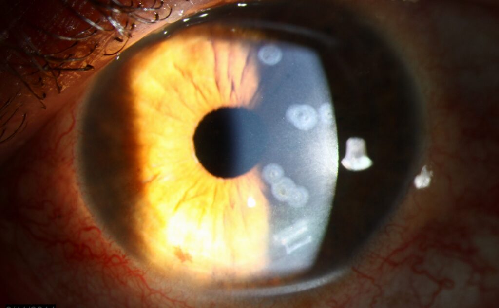
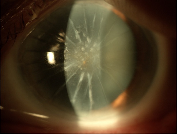
Corneal scarring is described in everyday practice with four classic terms that are short, easy to remember, and useful in notes and referral letters: nebula, macula, leucoma, and adherent leucoma.
They are graded by how dense the scar is and by whether the iris can be seen through it.
The four types
Nebula
A faint, superficial haze, visible only with the slit lamp or with oblique illumination.
It does not obscure the iris, and it usually causes little or no visual loss.
It is typically what is left after a mild superficial injury or a healed small infection.
Macula
A denser, more definite opacity that is visible without special illumination, for example, with a pen torch.
The iris details are still visible through it, but not clearly.
It causes some visual loss depending on its position.
Leucoma
A dense white scar that obscures the iris details and the pupil.
It results from full-thickness stromal damage.
If it is central, it causes severe visual loss.
Adherent leucoma
A leucoma in which the iris is stuck to the back of the scar, after a perforation.
The iris is incarcerated in the scar, and the anterior chamber is often shallow, with peripheral anterior synechiae.
This form has a risk of secondary glaucoma.
What causes them
- Infectious keratitis, bacterial, fungal, viral, and Acanthamoeba (see Wessely ring for one stromal immune sign)
- Trauma, including chemical injury and penetrating wounds
- Previous corneal surgery and complications
- Inflammation, such as herpetic stromal keratitis
- Nutritional deficiencies such as vitamin A deficiency in children (see Bitot spots)
- Trachoma and other cicatrizing diseases (see trachoma)
Corneal scarring is one of the leading causes of blindness worldwide, particularly in low-income countries where infection and trauma are common.
Fundus Explorer Pro
Photograph the retinal findings described here with the phone already in your pocket — 22 D optics and built-in illumination in one handheld unit.
From Choroida — the team behind this siteHow to assess a scar
- Visual acuity, with pinhole and refraction, to check how much of the loss is due to the scar and how much is irregular astigmatism
- Slit-lamp examination for depth, density, vascularization, and relation to the pupil
- Corneal thickness and topography, to see whether the cornea is thin or irregular
- Anterior segment OCT for depth of the scar (see anterior segment OCT)
- Assessment of the angle, pressure, and the posterior segment when the view allows (B-scan if not)
Treatment options
The right treatment depends on the depth, location, and visual need.
- Observation for peripheral or faint scars
- Contact lenses, including rigid gas-permeable or scleral lenses, can improve vision by masking irregularity (see scleral contact lens fitting)
- Phototherapeutic keratectomy for superficial scars
- Lamellar keratoplasty, where scars are anterior and the endothelium is healthy (see deep anterior lamellar keratoplasty)
- Penetrating keratoplasty for full-thickness scars
- Keratoprosthesis in eyes with repeated graft failure or severe surface disease (see Boston keratoprosthesis)
- Corneal tattooing for cosmetic purposes in blind eyes
In adherent leucoma, glaucoma control and careful surgical planning are required.
Examining a scar
Measure the size, depth, and location of the scar, and note the vascularization and the thickness of the cornea around it. Check the visual acuity with pinhole and with a contact lens trial, which can indicate how much improvement is possible by correcting irregular astigmatism. Examine the angle, pressure, and lens, and attempt to see the fundus. A B-scan is needed when the view is blocked.
Matching the treatment to the scar
Superficial scars in a quiet eye are the best candidates for phototherapeutic keratectomy. Deeper anterior scars with a healthy endothelium suit deep anterior lamellar keratoplasty, which avoids the risk of endothelial rejection. Full-thickness scars, or those with endothelial disease, need penetrating or endothelial-sparing combined procedures. Eyes with severe surface disease, repeated graft failure, or extensive vascularization may need a keratoprosthesis.
Prevention of corneal blindness
Many scars could be prevented by prompt treatment of infection, safe handling of chemicals, eye protection at work, vitamin A supplementation where deficiency occurs, and better access to eye care. Educating patients to seek help early for a red, painful eye is one of the most effective measures in clinical practice.
Documenting the scar
Photograph the scar and record its size, depth, and location. Note the visual acuity with and without correction. This information is useful to compare over time and to plan surgery, and it avoids confusion if the patient moves between clinics.
Counselling about visual expectations
A patient with a dense central scar should be told what to expect from each option, including the chance of improvement and the risk of graft failure. When the other eye is healthy, vision may be good enough for daily life, and some patients prefer to avoid surgery.
Prognosis
A nebula usually needs no treatment.
Visual outcomes after transplantation depend on the cause, vascularization, and ocular surface health, and are best for scars in otherwise quiet, avascular corneas.


Document what you see
Two smartphone imaging tools built for everyday clinic use — one for the slit lamp, one for the fundus.
From Choroida — the team behind this siteReferences
- Whitcher JP, Srinivasan M, Upadhyay MP. Corneal blindness: a global perspective. Bull World Health Organ. 2001;79:214-221.
- Gain P, Jullienne R, He Z, et al. Global survey of corneal transplantation and eye banking. JAMA Ophthalmol. 2016;134:167-173.
- Kanski JJ, Bowling B. Clinical Ophthalmology: A Systematic Approach. 7th ed. London: Elsevier; 2011.
- Krachmer JH, Mannis MJ, Holland EJ, eds. Cornea. 3rd ed. Philadelphia: Elsevier; 2011.
Test yourself
A few questions straight from this article.
-
A faint superficial corneal haze seen only on slit lamp, not obscuring the iris, is called what?
A nebula is a faint superficial haze visible only with the slit lamp or oblique illumination; it does not obscure the iris and usually causes little or no visual loss. -
Which corneal scar is visible with a pen torch while iris details remain visible but unclear?
A macula is a denser, definite opacity visible without special illumination; iris details can still be seen through it, though not clearly, and it causes some visual loss depending on position. -
A corneal leucoma results from damage of which extent?
A leucoma is a dense white scar from full-thickness stromal damage that obscures the iris details and the pupil, causing severe visual loss when central. -
What defines an adherent leucoma?
An adherent leucoma follows a perforation: the iris is stuck to the back of the scar and incarcerated in it, with a shallow anterior chamber and peripheral anterior synechiae. -
Which complication is specifically associated with an adherent leucoma?
Adherent leucoma carries a risk of secondary glaucoma, so glaucoma control and careful surgical planning are required. -
Why is acuity checked with a pinhole when assessing a corneal scar?
Pinhole acuity and refraction show how much of the visual loss comes from the opacity itself and how much from irregular astigmatism, which a contact lens trial can correct. -
Which procedure suits a superficial corneal scar in an otherwise quiet eye?
Superficial scars in a quiet eye are the best candidates for phototherapeutic keratectomy, while deeper and full-thickness scars need lamellar or penetrating grafts. -
Why is deep anterior lamellar keratoplasty preferred for a deep anterior scar with a healthy endothelium?
Deeper anterior scars with a healthy endothelium suit deep anterior lamellar keratoplasty, which spares the host endothelium and so avoids endothelial rejection. -
Which eyes with corneal scarring may require a keratoprosthesis?
A keratoprosthesis is reserved for eyes with repeated graft failure, severe ocular surface disease or extensive vascularization. -
Visual outcomes after corneal transplantation for scarring are best in which corneas?
Outcomes after transplantation depend on the cause, the degree of vascularization and ocular surface health, and are best for scars in otherwise quiet, avascular corneas.

Writes and reviews the clinical articles on Educate. Every post is checked against peer-reviewed sources before it goes live.


