A single infection, repeated over and over across childhood, can quietly reshape the inside of an eyelid until it turns the eye’s own lashes into the enemy.
Trachoma is the world’s leading infectious cause of blindness, and it does its damage slowly, over years of repeated infection.
It is entirely preventable and treatable, yet it remains a public health problem in the poorest, most water-scarce communities on earth.
The disease follows a well-defined progression, from simple follicular conjunctivitis in early childhood to scarring, inward-turned lashes, and corneal blindness decades later.
Recognizing where a patient sits on that spectrum is what determines whether antibiotics or eyelid surgery is the right next step.
Understanding trachoma’s staged natural history is essential for anyone working in regions where it remains endemic.
What Is Trachoma?
Trachoma is a chronic, recurrent conjunctival infection caused by Chlamydia trachomatis serotypes A-C, and it is graded using a simplified WHO staging system:
- Trachomatous inflammation, follicular (TF) — follicles on the upper tarsal conjunctiva, the hallmark of active disease
- Trachomatous inflammation, intense (TI) — pronounced inflammatory thickening obscuring the deep tarsal vessels
- Trachomatous scarring (TS) — visible scarring of the tarsal conjunctiva from repeated infection
- Trachomatous trichiasis (TT) — at least one lash rubbing on the globe, from scar-induced eyelid distortion
- Corneal opacity (CO) — visible corneal scarring, the end-stage blinding complication
This staging system exists because trachoma is fundamentally a disease of cumulative scarring — a single infection rarely blinds anyone; repeated infection over years does.
Epidemiology
Trachoma remains the leading infectious cause of blindness worldwide, concentrated in specific high-risk regions.
- It is endemic in parts of Africa, the Middle East, Central and South Asia, and some regions of Latin America and Australia
- Active disease (TF) is most common in preschool-aged children, who serve as the main reservoir for ongoing transmission
- Blinding complications (TT and CO) present decades later, predominantly in adult women, who have more contact with infected children as caregivers
- Prevalence is closely tied to water scarcity, poor sanitation, and overcrowding
The gap between childhood infection and adult blindness — often twenty to thirty years — is central to understanding why sustained, community-level control programs are needed rather than one-time treatment campaigns.
All-fit Slit-Lamp Adapter
Record and share exactly what you see at the slit lamp. One adapter fits any slit lamp or surgical microscope — and any smartphone.
From Choroida — the team behind this sitePathophysiology
Trachoma’s damage accumulates through a repeated cycle of infection and healing.
- Chlamydia trachomatis infects the conjunctival epithelium, provoking a follicular and papillary inflammatory response
- Repeated reinfection, common in young children in endemic communities, drives chronic inflammation rather than a single self-limited episode
- Chronic inflammation triggers fibrosis and progressive scarring of the tarsal conjunctiva over years
- Conjunctival scarring distorts the eyelid architecture, eventually turning the lashes inward (trichiasis)
- Constant mechanical trauma from misdirected lashes abrades the cornea, leading to corneal scarring and opacification
Because it is the cumulative scarring from repeated infections, not any single episode, that ultimately blinds, breaking the cycle of reinfection in early childhood is the entire basis of trachoma control.
Risk Factors
Environmental and Socioeconomic Risk Factors
- Limited access to clean water for face and hand washing
- Poor sanitation and limited access to latrines, which increases fly populations that transmit infection
- Overcrowded living conditions
- Low general awareness of hygiene practices that reduce transmission
Individual Risk Factors
- Young age, given the highest rates of active infection in preschool children
- Frequent close contact with infected children, a particular risk for female caregivers who go on to develop late complications
- Living in a household or community with high rates of active disease
Because transmission is driven by close contact and poor hygiene infrastructure rather than individual behavior alone, control requires community-wide intervention, not just treating individual patients.
Clinical Presentation
Symptoms
- Mild irritation, redness, and discharge in active childhood disease, often with few symptoms severe enough to prompt care
- Foreign-body sensation and pain once trichiasis develops in adulthood
- Progressive, painless vision loss as corneal opacification advances
Examination Findings
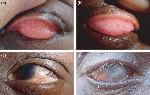
- Five or more follicles on the everted upper tarsal conjunctiva in active disease (TF)
- Diffuse inflammatory thickening obscuring the normal deep tarsal vessels in more intense disease (TI)
- Visible white, linear or stellate scarring of the tarsal conjunctiva in longstanding disease (TS)
- Inward-turned eyelashes touching the globe (TT), with or without associated entropion
- Corneal haze or opacity, sometimes with associated pannus, in end-stage disease (CO)
Everting the upper eyelid to examine the tarsal conjunctiva is the single most important step — none of these findings are visible without it.
Diagnostic Evaluation
Clinical Grading
- Diagnosis and staging are primarily clinical, using the WHO simplified grading system (TF, TI, TS, TT, CO)
- Systematic eyelid eversion in every at-risk patient, particularly in community screening programs
Laboratory Confirmation
- PCR testing for Chlamydia trachomatis can confirm active infection where available, though diagnosis in endemic settings is usually clinical
- Laboratory confirmation is more relevant for research, surveillance, and monitoring elimination progress than for routine individual case management
In endemic, resource-limited settings, the WHO grading system by itself — not laboratory testing — is what actually drives both individual treatment and community intervention decisions.
Differential Diagnosis
Conditions that can resemble trachoma include:
- Other causes of follicular conjunctivitis — viral (adenoviral) conjunctivitis, molluscum contagiosum, and toxic follicular reactions to topical medications
- Vernal keratoconjunctivitis — giant papillae rather than follicles, and a strong allergic/atopic history
- Ocular cicatricial pemphigoid — progressive conjunctival scarring without the geographic, contact-driven epidemiology of trachoma
- Other causes of entropion and trichiasis — age-related or post-traumatic, without the characteristic tarsal scarring pattern
Geographic context and exposure history carry substantial diagnostic weight — trachoma is overwhelmingly a disease of specific endemic regions rather than a random differential to consider everywhere.
Management
The WHO SAFE strategy structures both individual treatment and community-level control:
Surgery
- Trichiasis surgery to reposition the eyelid margin and stop lashes from touching the cornea, preventing progression to blindness
- Surgery is the priority intervention for anyone already in the TT stage, since it directly prevents further corneal damage
Antibiotics
- Single-dose oral azithromycin is the treatment of choice for active infection, both for individuals and for mass community treatment in endemic areas
- Community-wide mass drug administration is used to reduce the overall burden of infection and interrupt transmission
Facial Cleanliness and Environmental Improvement
- Promotion of regular face washing to reduce transmission, particularly among young children
- Improved access to water and sanitation infrastructure, along with fly control, to reduce community-level transmission over the long term
Surgery treats the immediate threat to vision; antibiotics and hygiene/environmental measures are what actually prevent the disease from recurring in the community.
Prognosis
Outcomes depend heavily on the stage at which disease is identified and treated.
- Active follicular disease (TF/TI) treated with antibiotics carries an excellent prognosis with no lasting damage
- Trichiasis caught and surgically corrected before significant corneal damage prevents progression to blindness
- Established corneal opacity (CO) is largely irreversible, and represents the cumulative result of years of untreated or repeated disease
Global elimination efforts have made real progress, but the decades-long lag between childhood infection and adult blindness means the full benefit of current control programs will not be seen for a generation.


Document what you see
Two smartphone imaging tools built for everyday clinic use — one for the slit lamp, one for the fundus.
From Choroida — the team behind this siteReferences
- World Health Organization. Trachoma. WHO Fact Sheets.
- Solomon AW, Zondervan M, Kuper H, et al. Trachoma control: a guide for programme managers. World Health Organization.
- Taylor HR, Burton MJ, Haddad D, et al. Trachoma. The Lancet. 2014.
- Hu VH, Harding-Esch EM, Burton MJ, et al. Epidemiology and control of trachoma: systematic review. Tropical Medicine & International Health. 2010.
- Trachoma. EyeWiki, American Academy of Ophthalmology.
Test yourself
A few questions straight from this article.
-
Which organism causes trachoma?
Trachoma is a chronic, recurrent conjunctival infection caused by Chlamydia trachomatis serotypes A-C, transmitted in crowded, water-scarce communities. -
In the WHO simplified grading of trachoma, what defines trachomatous inflammation, follicular (TF)?
TF, the hallmark of active trachoma, is graded by finding five or more follicles on the everted upper tarsal conjunctiva. Thickening that obscures the deep tarsal vessels defines the more intense grade, TI. -
In the WHO grading of trachoma, what defines trachomatous trichiasis (TT)?
TT is graded when at least one eyelash rubs on the globe, the result of scar-induced eyelid distortion and the point at which surgery becomes the priority intervention. -
What is trachoma's global public health significance?
Trachoma remains the world's leading infectious cause of blindness, endemic in parts of Africa, the Middle East, Central and South Asia, and some regions of Latin America and Australia. -
In endemic areas, which group most often presents with the blinding complications of trachoma?
Active follicular disease is commonest in preschool children, who are the transmission reservoir, but trichiasis and corneal opacity appear decades later, predominantly in the adult women who care for them. -
What directly causes the corneal scarring that blinds patients with trachoma?
Years of repeated infection scar the tarsal conjunctiva until the eyelid distorts and the lashes turn inward; constant mechanical trauma from those lashes abrades the cornea into opacity. -
Which examination step is essential to diagnose and grade trachoma?
Every WHO grading sign sits on the upper tarsal conjunctiva, so systematic eversion of the upper eyelid is the single indispensable step in both clinic and community screening. -
What is the antibiotic treatment of choice for active trachoma infection?
Single-dose oral azithromycin is the treatment of choice, used both for individual patients and for community-wide mass drug administration to interrupt transmission. -
Within the WHO SAFE strategy, which intervention is the priority for a patient already at the trichiasis stage of trachoma?
Surgery is prioritised at the TT stage because repositioning the eyelid margin stops the lashes touching the cornea and directly prevents progression to blindness. -
Which stage of trachoma is largely irreversible?
Established corneal opacity is largely irreversible and represents years of untreated, repeated disease, whereas active follicular disease treated with antibiotics leaves no lasting damage.

Writes and reviews the clinical articles on Educate. Every post is checked against peer-reviewed sources before it goes live.