
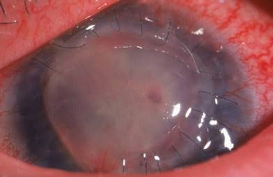
Corneal wound dehiscence is a separation or opening of a previously closed corneal or limbal surgical wound, occurring either in the early postoperative period from inadequate initial closure or, less commonly, years later from trauma or spontaneous wound failure.
It is a genuine ophthalmic emergency in most cases, since an open wound risks intraocular content prolapse, infection, and severe, sometimes irreversible visual loss if not recognized and repaired promptly.
Understanding both the acute postoperative form and the delayed, sometimes trauma-related form clarifies why any corneal or limbal surgical wound remains a point of potential vulnerability for the rest of a patient’s life, not just during the immediate recovery period.
Modern small-incision techniques have reduced the overall frequency of this complication compared to the era of larger, sutured incisions, but the underlying principle of lifelong relative wound weakness has not changed.
Causes
- Inadequate initial wound construction or closure during the original surgery, including a poorly angled clear corneal incision from phacoemulsification, discussed in its own dedicated article on this site, that fails to seal properly
- Premature suture removal or spontaneous suture loosening or breakage after a sutured procedure such as penetrating keratoplasty, discussed in its own dedicated article on this site
- Blunt or penetrating trauma to an eye with a prior surgical wound, which tends to fail preferentially at the site of that old, structurally weaker scar rather than through healthy, unoperated tissue
- Elevated intraocular pressure or vigorous eye rubbing in the early postoperative period, placing mechanical stress on a wound that has not yet achieved full structural strength
Why Old Surgical Wounds Remain Vulnerable
A corneal or limbal surgical wound never regains the full tensile strength of unoperated corneal tissue, even years after apparently complete healing, since the scar tissue that forms is structurally different from native, undisturbed stroma.
This persistent, lifelong relative weakness is why a patient with any history of penetrating keratoplasty, cataract surgery, or other corneal or limbal incisional surgery is at somewhat elevated risk of wound dehiscence specifically at that old surgical site if the eye later sustains blunt trauma, even trauma that might cause only minor injury to an eye without a surgical history.
Patients with a significant surgical history benefit from understanding this ongoing vulnerability, particularly around activities carrying meaningful risk of blunt ocular trauma, so that appropriate protective eyewear can be considered where relevant.
All-fit Slit-Lamp Adapter
Record and share exactly what you see at the slit lamp. One adapter fits any slit lamp or surgical microscope — and any smartphone.
From Choroida — the team behind this siteClinical Presentation
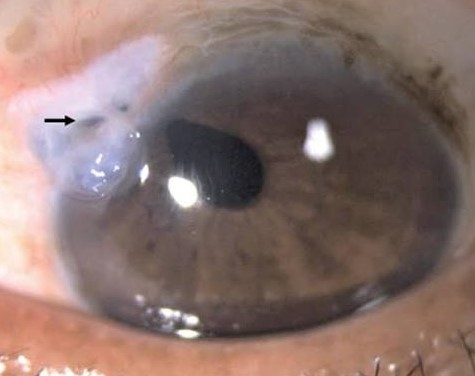
Acute postoperative dehiscence typically presents with sudden pain, decreased vision, and a shallow or flat anterior chamber, sometimes with visible wound gape, iris prolapse, or frank leakage of aqueous humor from the wound.
Delayed, trauma-related dehiscence presents similarly following an injury, and the history of prior surgery at the affected site, even if remote, should immediately raise suspicion for wound rupture rather than a simple contusion injury.
A positive Seidel test, showing aqueous leakage as a streaming, diluted line through instilled fluorescein dye, confirms an open wound and active leak when the diagnosis is uncertain on gross exam alone.
Evaluation
Careful slit-lamp examination assesses the extent of wound separation, anterior chamber depth, and whether any intraocular content, including iris, lens material, or vitreous, has prolapsed through the open wound.
Imaging, including ultrasound when a view to the posterior segment is obscured, helps assess for associated posterior segment injury, particularly important in a case involving trauma rather than a purely spontaneous postoperative wound failure.
Assessment should proceed cautiously and gently, avoiding any additional pressure on the globe, given the risk of extruding further intraocular content through an already compromised, open wound during the examination itself.
Management
Prompt surgical repair in the operating room is required for any significant wound dehiscence, resuturing the wound and addressing any prolapsed tissue, whether by repositioning viable tissue or excising tissue that is no longer viable.
Broad-spectrum antibiotic coverage is initiated given the meaningful infection risk associated with an open ocular wound, following similar urgency and principles to management of a ruptured globe from any other cause.
Postoperative management after repair mirrors that of the original surgery in many respects, with particular attention to protecting the newly repaired wound from any further mechanical stress during the critical early healing period.
Visual outcome after successful repair depends heavily on how much intraocular content was lost or damaged before repair could be performed, which is one more reason prompt recognition and same-day surgical repair matter as much as the technical quality of the repair itself.


Document what you see
Two smartphone imaging tools built for everyday clinic use — one for the slit lamp, one for the fundus.
From Choroida — the team behind this siteReferences
- American Academy of Ophthalmology. Basic and Clinical Science Course, Section 8: External Disease and Cornea.
- Kloek CE, Bordón AF, Chen TC, Pineda R II. Wound dehiscence after penetrating keratoplasty. Seminars in Ophthalmology.
- Bowling B. Kanski’s Clinical Ophthalmology.
Test yourself
A few questions straight from this article.
-
What defines corneal wound dehiscence?
Dehiscence is the separation or opening of a previously closed corneal or limbal surgical wound, either early after surgery from inadequate closure or years later from trauma or spontaneous failure. -
Why does a corneal surgical wound remain vulnerable years after apparently complete healing?
The scar that forms is structurally different from undisturbed stroma and never regains full tensile strength, leaving a lifelong relative weakness at the old surgical site. -
Where does an eye with previous corneal surgery preferentially fail after blunt trauma?
Trauma to an eye with a prior surgical wound tends to fail preferentially at the structurally weaker old scar rather than through healthy, unoperated tissue. -
Which anterior chamber finding accompanies acute postoperative corneal wound dehiscence?
Acute dehiscence typically presents with sudden pain, reduced vision and a shallow or flat anterior chamber, sometimes with visible wound gape, iris prolapse or frank aqueous leakage. -
What does a positive Seidel test demonstrate in a suspected corneal wound dehiscence?
A positive Seidel test shows aqueous streaming as a diluted line through instilled fluorescein, confirming an open wound with an active leak when gross examination is inconclusive. -
How should the eye be examined when corneal wound dehiscence is suspected?
Examination must be gentle and avoid pressure on the globe, because further intraocular content can be extruded through an already compromised open wound during the assessment itself. -
Which investigation helps assess posterior segment injury when the fundus view is obscured?
When the posterior segment cannot be seen, ultrasound helps assess for associated posterior injury, which matters particularly in trauma-related rather than purely postoperative wound failure. -
What is the required treatment for a significant corneal wound dehiscence?
Significant dehiscence needs prompt theatre repair, resuturing the wound and either repositioning viable prolapsed tissue or excising tissue that is no longer viable. -
Why is broad-spectrum antibiotic cover started in corneal wound dehiscence?
An open ocular wound carries a meaningful infection risk, so broad-spectrum cover is started following the same urgency and principles as a ruptured globe from any other cause. -
What most strongly determines visual outcome after repair of corneal wound dehiscence?
Outcome depends heavily on how much intraocular content was lost or damaged before repair could occur, which is why prompt recognition and same-day surgery matter as much as technical quality.

Writes and reviews the clinical articles on Educate. Every post is checked against peer-reviewed sources before it goes live.


