
CASE REPORT
A 45-year-old man was referred to the ocular oncology service for evaluation due to a black asymptomatic lesion in his right eye. His medical history included hyperlipidemia and high myopia.
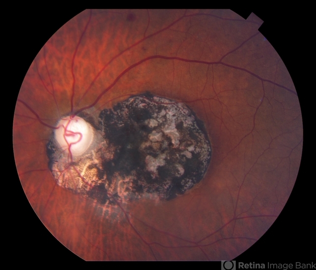
On clinical examination, a flat black lesion that looked like a flower was observed. The lesion did not resemble melanoma. Moreover, an ultrasound examination confirmed that the lesion was flat.
The lesion showed hypo autofluorescence at the dark parts that correlate with the known histopathologic evidence of lack of lipofuscin in the retinal pigment epithelium of congenital hypertrophy of retinal pigment epithelium (CHRPE) and mild hyper autofluorescence that correlate with scleral autofluorescence at the lacunae.
In optical coherence tomography, a loss of the external retinal layers was seen in the area of the lesion and no retina was seen in the lacunae’ area. The diagnosis was confirmed as Congenital Hypertrophy of the Retinal Pigment Epithelium (CHRPE).
Congenital Hypertrophy of the Retinal Pigment Epithelium (CHRPE) DISEASE entity
Congenital hypertrophy of the retinal pigment epithelium (CHRPE) is a typically benign, asymptomatic, pigmented fundus lesion. It is a congenital hamartoma of the retinal pigment epithelium (RPE) and occurs in three variant forms: solitary (unifocal), grouped (multifocal), and atypical.
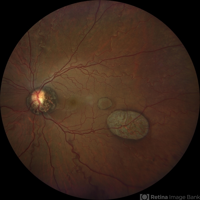
Atypical CHRPE is associated with familial adenomatous polyposis (FAP), an autosomal dominant cancer syndrome, characterized by numerous adenomatous polyps of the colon and rectum. Left untreated, virtually all FAP patients develop colorectal carcinoma/s by middle age.
FAP sub-types, including Gardner syndrome (FAP plus skeletal hamartomas and various soft tissue tumors) and Turcot syndrome (FAP plus various brain tumors), are also associated with atypical CHRPE.
The prevalence of CHRPE in the general optometric population has been estimated to be 1.2%. Atypical CHRPE is the earliest and most common extra-colonic manifestation of FAP, present in up to 90% of patients.
Congenital Hypertrophy of the Retinal Pigment Epithelium (CHRPE) Diagnosis
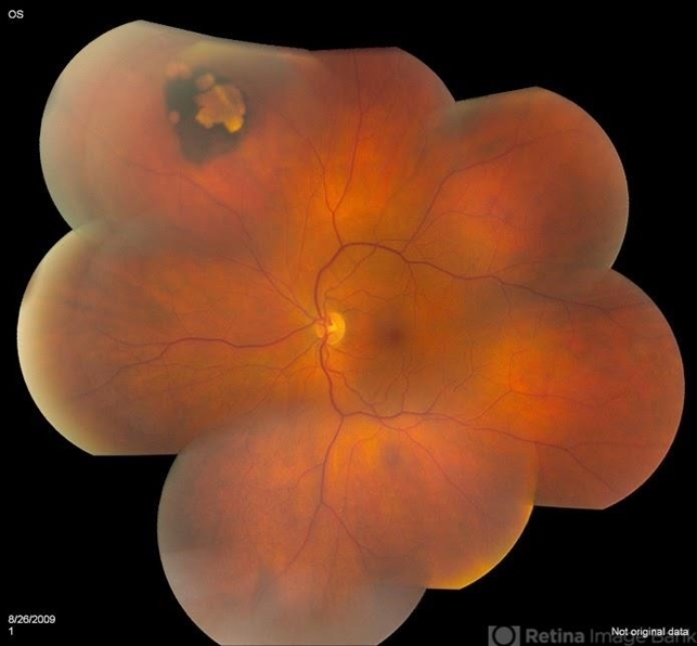
The diagnosis of CHRPE is usually made clinically and no diagnostic procedures are generally necessary. Fundus photography is useful for documentation and follow-up of lesions and wide-field scanning-laser ophthalmoscopy has been recommended as a screening tool.
Ancillary testing may be beneficial in uncertain cases.
- Lesions typically demonstrate hypo autofluorescence on fundus autofluorescence (FAF) due to their high melanin content. Non-pigmented haloes or lacunae may show autofluorescence.
- No leakage is demonstrated on fluorescein angiography (FA) or indocyanine green angiography (ICGA). Lesions typically block underlying choroidal fluorescence, except in areas of depigmented lacunae or haloes.
- Optical coherence tomography findings include retinal thinning and photoreceptor loss over lesions, with the absence of RPE and increased transmission of light in areas of lacunae.
- The use of Optical coherence tomography angiography (OCTA) in the evaluation of CHRPE has limitations due to thick RPE and increased cumulative melanin granules. However, this modality is better than FA or ICG in visualizing choroidal vasculature.
- Electroretinogram (ERG), electrooculogram (EOG), A-scan, and B-scan ultrasonography are non-contributory.
Fundus Explorer Pro
Photograph the retinal findings described here with the phone already in your pocket — 22 D optics and built-in illumination in one handheld unit.
From Choroida — the team behind this siteDifferential diagnosis
- Choroidal melanoma
- Choroidal nevus
- Melanocytoma
- Focal pigmentation (caused by injury, inflammation, or drug toxicity)
- True hyperplasia of the RPE
- Black sunburst lesion in sickle cell retinopathy
- Congenital grouped albinotic retinal pigment epithelial spots (CGARPES) (“polar bear tracks”) may resemble grouped CHRPE but are characterized by multiple grouped, white, variably-sized, albinotic spots of the RPE. The nature of these lesions has not been investigated histologically.
MANAGEMENT of Congenital Hypertrophy of the Retinal Pigment Epithelium (CHRPE)
No active intervention is generally indicated or required. Proton beam therapy has been described for rare complicated cases.
Complications
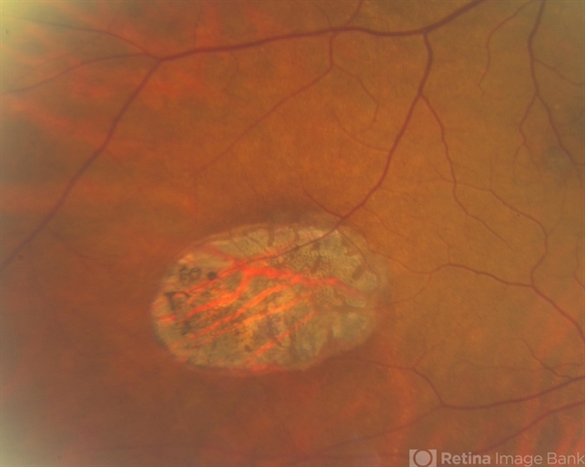
CHRPE lesions have been documented to enlarge in 46-83% of cases over at least three years of follow-up. The foveal extension may result in impaired visual acuity.
Rarely, nodular pigmented adenocarcinomas may arise from within areas of CHRPE. Untreated nodular lesions have been documented to progress to pedunculated tumors with associated serous retinal detachment.
Premacular gliosis and cystoid macular edema are frequently seen when peripheral pigmented or non-pigmented RPE tumors arise within CHRPE.


Document what you see
Two smartphone imaging tools built for everyday clinic use — one for the slit lamp, one for the fundus.
From Choroida — the team behind this siteREFERENCES
- Coleman P, Barnard NA. Congenital hypertrophy of the retinal pigment epithelium: prevalence and ocular features in the optometric population. Ophthalmic Physiol Opt. 2007;27:547–555.
- Wallis YL, Macdonald F, Hulten M, et al: Genotype-phenotype correlation between position of constitutional APC gene mutation and CHRPE expressed in FAP. Hum Gene. 1994;94:543-548.
- Traboulsi ET, Apostoides J, Giardiello FM, et al. Pigmented ocular fundus lesions and APC mutations in familial adenomatous polyposis. Ophth Genet. 1996;17:167-174.
- Deibert B, Ferris L, Sanchez N, Weishaar P. The link between colon cancer and congenital hypertrophy of the retinal pigment epithelium (CHRPE). Am J Ophthalmol Case Rep. 2019 Sep;15:100524.
- Nieuwenhuis MH, Vasen HF. Correlations between mutation site in APC and phenotype of familial adenomatous polyposis (FAP): a review of the literature. Crit Rev Oncol Hematol. 2007;61:153-161.
Test yourself
A few questions straight from this article.
-
What kind of lesion is congenital hypertrophy of the retinal pigment epithelium (CHRPE)?
CHRPE is a congenital hamartoma of the RPE and is typically a benign, asymptomatic pigmented fundus lesion. -
In which three variant forms does CHRPE occur?
CHRPE is described in three variants: solitary (unifocal), grouped (multifocal) and atypical. -
Which CHRPE variant is associated with familial adenomatous polyposis?
Atypical CHRPE is the variant linked to familial adenomatous polyposis, an autosomal dominant cancer syndrome, and to its Gardner and Turcot subtypes. -
What is the estimated prevalence of CHRPE in the general optometric population?
CHRPE prevalence in the general optometric population has been estimated at 1.2%. -
In up to what proportion of familial adenomatous polyposis patients is atypical CHRPE present?
Atypical CHRPE is the earliest and most common extra-colonic manifestation of familial adenomatous polyposis, present in up to 90% of patients. -
What is the typical fundus autofluorescence appearance of a CHRPE lesion?
Dense melanin makes CHRPE hypoautofluorescent on FAF, while non-pigmented haloes or lacunae within the lesion may show autofluorescence. -
Which entity resembles grouped CHRPE but consists of white albinotic spots known as polar bear tracks?
CGARPES produces multiple grouped, white, variably sized albinotic RPE spots that can mimic grouped CHRPE despite being pale rather than pigmented. -
Which OCT feature is seen within the lacunae of a CHRPE lesion?
Over the lesion OCT shows retinal thinning and photoreceptor loss, while in the lacunae the RPE is absent and light transmission is increased. -
Which rare malignant complication can arise from within an area of CHRPE?
Nodular pigmented adenocarcinomas rarely arise within CHRPE, and untreated nodular lesions have progressed to pedunculated tumours with serous retinal detachment. -
What is the standard management of an uncomplicated CHRPE lesion?
No active treatment is generally indicated; fundus photography documents and monitors the lesion, and proton beam therapy is reserved for rare complicated cases.
