
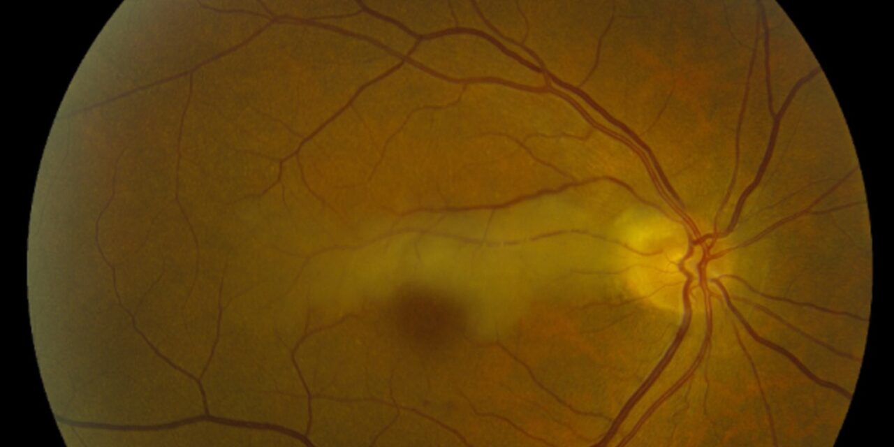
Cilioretinal artery occlusion is a blockage of the cilioretinal artery, a small vessel present in a minority of eyes that arises from the choroidal circulation rather than the central retinal artery.
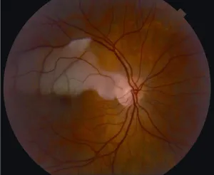
Because it has a separate origin from the central retinal artery, its occlusion produces a visual and fundus picture genuinely different from a typical central or branch retinal artery occlusion, and the two should not be lumped together diagnostically.
Whether central vision is spared or devastated by this occlusion depends almost entirely on one anatomic detail: whether the cilioretinal artery happens to supply the papillomacular bundle.
Cilioretinal artery occlusion is uncommon on its own, but it shows up in three distinct clinical contexts that are worth keeping separate: an isolated occlusion, a combined occlusion alongside central retinal vein occlusion, and, occasionally, as a presenting feature of giant cell arteritis.
Anatomy
A cilioretinal artery is present in a substantial minority of eyes, typically emerging from the temporal edge of the optic disc and supplying a variable wedge of retina between the disc and the macula.
Because it branches from the posterior ciliary circulation rather than the central retinal artery, it is not affected when the central retinal artery itself becomes occluded, which is why some patients with a central retinal artery occlusion retain a small island of preserved central vision.
The clinical significance of the cilioretinal artery, in other words, only becomes apparent when either it or the central retinal artery is blocked, since in a normal eye its contribution is not something a patient or examiner would ever notice.
Presentation as an Isolated Occlusion
When the cilioretinal artery is occluded on its own, with the central retinal artery still patent, the visual outcome depends heavily on whether the affected wedge of retina includes the papillomacular bundle.
If the papillomacular bundle is spared, the patient may notice only a paracentral scotoma with preserved, sometimes near-normal, central acuity.
If the papillomacular bundle lies within the cilioretinal artery’s territory, an isolated occlusion can cause a dense central scotoma every bit as devastating as a typical central retinal artery occlusion, despite the fact that only a single small branch vessel is involved.
Fundus Explorer Pro
Photograph the retinal findings described here with the phone already in your pocket — 22 D optics and built-in illumination in one handheld unit.
From Choroida — the team behind this sitePresentation Combined With Central Retinal Vein Occlusion
Cilioretinal artery occlusion is also seen as a combined finding with central retinal vein occlusion, a recognized and distinct clinical entity from either process occurring alone.
In this combined picture, venous congestion from the vein occlusion is thought to compromise perfusion through the watershed cilioretinal circulation, producing an arterial infarct superimposed on the venous changes.
Visual prognosis in this combined form tends to be worse than an uncomplicated central retinal vein occlusion alone, reflecting the added ischemic insult from the arterial component.
Fundus Findings
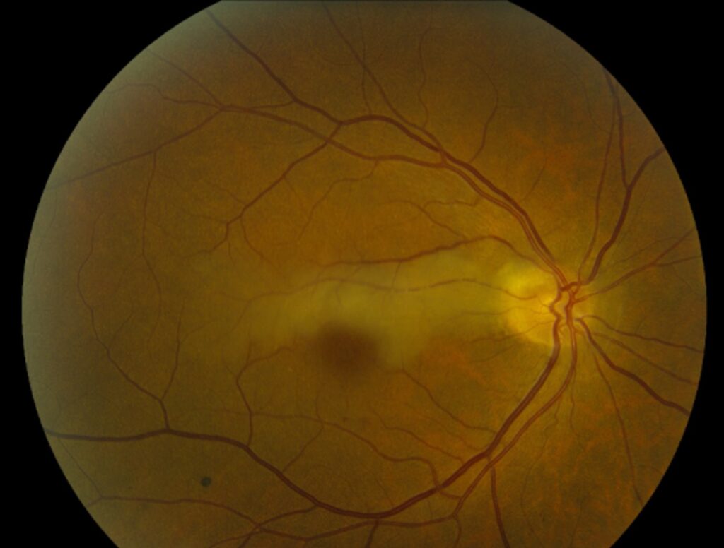
- A wedge or triangular area of retinal whitening extending from the optic disc toward the macula, corresponding to the cilioretinal artery’s distribution
- A relatively normal-appearing retina outside this wedge when the occlusion is isolated, in contrast to the diffuse whitening of a full central retinal artery occlusion
- Signs of the associated central retinal vein occlusion, when present, including diffuse hemorrhages and venous dilation and tortuosity
- A cherry-red spot may or may not be visible depending on whether the papillomacular bundle and foveal region fall within the ischemic wedge
Workup
The systemic workup mirrors that of any other acute retinal arterial occlusion, since the underlying causes overlap substantially: embolic disease from the carotids or heart, and, in an older patient, giant cell arteritis must be actively excluded given the risk to the fellow eye.
Giant cell arteritis deserves particular attention in a patient over 50 with new visual symptoms and any systemic features such as jaw claudication, scalp tenderness, or an elevated inflammatory marker, since cilioretinal artery occlusion has been described as a presenting feature of arteritic disease.
Fluorescein angiography can help confirm the diagnosis and define the extent of the occluded territory when the diagnosis is not entirely clear on clinical exam alone.
Optical coherence tomography typically shows inner retinal hyperreflectivity and thickening confined to the affected wedge in the acute phase, evolving into localized inner retinal thinning as the affected territory atrophies over the following weeks.
Management
There is no proven treatment that reliably reverses vision loss once cilioretinal artery occlusion has occurred, and management follows the same general framework used for other acute retinal arterial occlusions: urgent evaluation for a treatable embolic source and, when giant cell arteritis is suspected, immediate high-dose corticosteroids to protect the fellow eye rather than to reverse the vision already lost.
Long-term follow-up focuses on secondary stroke and cardiovascular risk reduction, since an acute retinal arterial occlusion of any type is a recognized marker of elevated systemic vascular risk.
When cilioretinal artery occlusion occurs alongside central retinal vein occlusion, management also has to address the venous component, including monitoring for the macular edema and neovascular complications that drive most of the visual morbidity in that combined presentation.


Document what you see
Two smartphone imaging tools built for everyday clinic use — one for the slit lamp, one for the fundus.
From Choroida — the team behind this siteReferences
- Hayreh SS, Podhajsky PA, Zimmerman MB. Central and hemicentral retinal vein occlusion: role of anti-platelet aggregation agents and anticoagulants. Ophthalmology.
- Brown GC, Duker JS, Lehman R, Eagle RC Jr. Combined central retinal artery-central vein obstruction. International Ophthalmology.
- American Academy of Ophthalmology. Basic and Clinical Science Course, Section 12: Retina and Vitreous.
- Hayreh SS. Acute retinal arterial occlusive disorders. Progress in Retinal and Eye Research.
Test yourself
A few questions straight from this article.
-
From which circulation does the cilioretinal artery arise?
The cilioretinal artery is present in a minority of eyes and branches from the choroidal circulation rather than the central retinal artery, which is why its occlusion behaves so differently. -
Why do some patients with central retinal artery occlusion retain a small island of central vision?
Because it branches from the posterior ciliary circulation, a cilioretinal artery is not affected when the central retinal artery occludes, and it can keep a wedge of central retina perfused. -
What determines whether an isolated cilioretinal artery occlusion spares central acuity?
If the papillomacular bundle is spared the patient may notice only a paracentral scotoma, but if it falls inside the occluded wedge the central scotoma can be as dense as in a central retinal artery occlusion. -
What is the classic fundus appearance of an acute cilioretinal artery occlusion?
The infarcted retina whitens in a wedge or triangle running from the optic disc toward the macula, matching the distribution the cilioretinal artery supplies. -
Which fundus feature separates an isolated cilioretinal artery occlusion from a full central retinal artery occlusion?
An isolated cilioretinal occlusion whitens only its own territory, in contrast with the diffuse whitening seen across the posterior pole in a complete central retinal artery occlusion. -
How is central retinal vein occlusion thought to precipitate a combined cilioretinal artery occlusion?
Congestion from the vein occlusion is thought to impair perfusion through the watershed cilioretinal circulation, producing an arterial infarct superimposed on the venous picture. -
How does visual prognosis in combined cilioretinal artery and central retinal vein occlusion compare with vein occlusion alone?
The combined form carries a poorer visual prognosis than an uncomplicated central retinal vein occlusion, reflecting the extra ischaemic damage from the arterial component. -
What does optical coherence tomography show acutely in cilioretinal artery occlusion?
Acutely the inner retina in the affected wedge is hyperreflective and thickened, and over the following weeks that territory atrophies into localised inner retinal thinning. -
In which patient with cilioretinal artery occlusion must giant cell arteritis be actively excluded?
Cilioretinal artery occlusion has been described as a presenting feature of arteritic disease, so systemic features and raised inflammatory markers in a patient over 50 demand active exclusion of giant cell arteritis. -
Why are immediate high-dose corticosteroids given when giant cell arteritis causes a cilioretinal artery occlusion?
No treatment reliably reverses the vision already lost, so urgent steroids in suspected giant cell arteritis are given to safeguard the fellow eye rather than to recover the affected one.
