
CASE REPORT
A 70-year-old man was referred to a clinic because of bilateral fundus lesions. He had a history of chronic kidney disease (CKD) and secondary hyperparathyroidism. Smartphone Fundoscopy showed a cluster of choroidal folds in the superotemporal extra-macular region OS.

Swept-source optical coherence tomography demonstrated ellipsoid zone disruption OD, retinal pigment epithelium undulation OS, dilated Haller layer veins OU, and central choroidal thickening OU and thinning of the overlying choroid due to scleral elevation OS.
Fluorescein angiography detected macular hyperfluorescence OD. Indocyanine green angiography demonstrated choroidal vascular hyperpermeability together with numerous scattered hypofluorescent lesions OU.
Fundus autofluorescence showed multiple hypoautofluorescent spots surrounded by hyperautofluorescent areas OD. Laser speckle flowgraphy exhibited choroidal blood flow reduction represented by a cold color pattern OU.
B-mode echography displayed hyperechoic solid lesions with acoustic shadowing and orbital computed tomography revealed high-density areas in the sclera, both of which were consistent with calcification. The patient was diagnosed with Sclerochoroidal Calcification.
Sclerochoroidal Calcification DISEASE entity
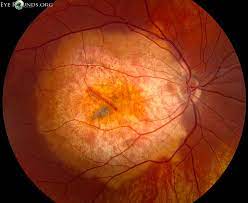
Sclerochoroidal calcification presents as flat or minimally elevated yellow placoid lesions, sometimes associated with gray lesions that are similar in appearance to senile scleral plaque. Sclerochoroidal Calcification is rare and frequently misdiagnosed.

The majority of cases are idiopathic and present with normal calcium-phosphorus metabolism and without associated ophthalmic or systemic conditions.
The majority of patients diagnosed with Sclerochoroidal Calcification are Caucasian and elderly. There is a slight female predisposition (approximately 60% of cases). Patients with previous systemic disorders that impact calcium-phosphorus metabolism are at higher risk of developing sclerochoroidal calcifications.
These include hyperparathyroidism, pseudohypoparathyroidism, parathyroid adenoma, chondrocalcinosis, sarcoidosis, Gitelman syndrome, Bartter syndrome, pseudo-gout, chronic renal disease, hypophosphatemia, hypervitaminosis, and metabolic imbalance after diuretic use.
Fundus Explorer Pro
Photograph the retinal findings described here with the phone already in your pocket — 22 D optics and built-in illumination in one handheld unit.
From Choroida — the team behind this siteSclerochoroidal Calcification Symptoms
Patients are often asymptomatic and do not report ocular complaints. Patients do not typically show a change in visual acuity. However, there are several case reports of macular SCC associated with choroidal neovascularization or exudative retinal detachment resulting in decreased visual acuity.
However, a large review of 179 eyes of 118 patients did not report hemorrhage, subretinal fluid, retinal pigment epithelial detachment, or choroidal neovascular membrane (CNVM) in any case.
A long follow-up (mean 4 years) of these cases did not show enlargement of the lesion, hemorrhage, subretinal fluid, CNVM, vision loss, or decalcification. None of the cases needed treatment.

Diagnostic Procedures
Ophthalmoscopy ( Smartphone Fundoscopy ) is sufficient for diagnosis. The fellow eye may also have a similar and symmetrical lesion. There may be multiple lesions which, however, are typically localized near the superotemporal arcade or at the equator.
There might be atrophy of the overlying choroid and/or retinal pigment epithelium. The lesions may be arranged in a ring-like configuration. Calcifications can also be detected by B-scan ultrasonography, optical coherence tomography, orbital computed tomography, fundus autofluorescence, fluorescein angiography, and indocyanine green angiography.
Ophthalmoscopy can be unremarkable while ultrasound shows echogenic areas indicative of calcifications (hyperechoic lesion with acoustic shadow), due to deposition in the sclera not yet reaching the choroid.
Sclerochoroidal Calcification MANAGEMENT
General Treatment
Parathyroid adenoma, Bartter, and Gitelman syndromes should be ruled out with testing due to complications associated with these diseases.
Patients should be advised to return annually for a dilated eye exam to monitor the progression of the lesions, although there is no treatment other than correcting any metabolic imbalances.
Prognosis
Calcifications are commonly misdiagnosed, resulting in unnecessary testing and referral to retinal specialists.
Lesions are typically located away from the macula and rarely encroach the fovea, therefore visual prognosis in patients is good and visual acuity usually remains unchanged.


Document what you see
Two smartphone imaging tools built for everyday clinic use — one for the slit lamp, one for the fundus.
From Choroida — the team behind this siteREFERENCES
- Honavar SG, Shields CL, Demirci H, & Shields JA. Sclerochoroidal calcification: clinical manifestations and systemic associations. Arch Ophthalmol. 2001;119(6), 833-840. doi:10.1001/archopht.119.6.833.
- Shields JA, Shields CL. CME Review: Sclerochoroidal calcification. Retina. 2002;22(3):251-261. doi:10.1097/00006982-200206000-00001.
- Shields CL, Hasanreisoglu M, Saktanasate J, Shields PW, Seibel I, Shields JA. Sclerochoroidal calcification. Retina. 2015;35(3):547-554. doi:10.1097/iae.0000000000000450.
- Thomson AC, Brown GT, Dolores-Rodriguez A, Hunter AA. A case of extensive bilateral idiopathic sclerochoroidal calcification and review of the literature. Int Med Case Rep J. 2021; 14:749-755. doi:10.2147/imcrj.s336237.
- Bourcier T, Blain P, Massin P, Grünfeld J-P, Gaudric A. Sclerochoroidal calcification associated with Gitelman syndrome. Am J Ophthalmol. 1999;128(6):767-768. doi:10.1016/s0002-9394(99)00277-9.
- Hasanreisoglu M, Saktanasate J, Shields PW, Shields CL. Classification of sclerochoroidal calcification based on enhanced depth imaging optical coherence tomography “mountain-like” features. Retina. 2015;35(7):1407-1414. doi:10.1097/iae.0000000000000468.
Test yourself
A few questions straight from this article.
-
How does sclerochoroidal calcification typically appear on fundus examination?
Sclerochoroidal calcification appears as flat or minimally elevated yellow placoid lesions, sometimes with grey areas; it is rare and frequently misdiagnosed. -
The grey lesions of sclerochoroidal calcification closely resemble which entity?
The grey lesions sometimes accompanying sclerochoroidal calcification look very like senile scleral plaque, one reason the condition is so often misdiagnosed. -
Where are the lesions of sclerochoroidal calcification typically located?
Lesions are typically near the superotemporal arcade or at the equator, may be multiple or ring-like, and the fellow eye often shows a symmetrical lesion. -
Which demographic group is most often affected by sclerochoroidal calcification?
Most patients are elderly and Caucasian, with a slight female predisposition accounting for roughly 60% of cases. -
What is true of calcium-phosphorus metabolism in most cases of sclerochoroidal calcification?
The majority of cases are idiopathic with normal calcium-phosphorus metabolism and no associated ocular or systemic disease, though metabolic disorders raise the risk. -
Which systemic conditions should be specifically excluded when sclerochoroidal calcification is found?
Parathyroid adenoma, Bartter syndrome and Gitelman syndrome should be ruled out by testing because of the complications associated with these diseases. -
What does B-scan ultrasonography show in sclerochoroidal calcification?
Echography shows hyperechoic solid lesions casting acoustic shadows, and orbital CT confirms high-density areas within the sclera. -
What did long-term follow-up of a large sclerochoroidal calcification series show?
Across 179 eyes of 118 patients followed for a mean of four years there was no enlargement, haemorrhage, subretinal fluid, neovascular membrane, vision loss or decalcification, and no case required treatment. -
How should a patient with sclerochoroidal calcification be followed and treated?
Patients return annually for a dilated examination to monitor lesion progression; there is no treatment beyond correcting any underlying metabolic imbalance. -
Why is the visual prognosis in sclerochoroidal calcification generally good?
Lesions are typically located away from the macula and rarely encroach on the fovea, so visual acuity usually remains unchanged.
