
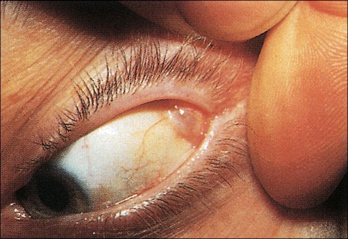
Lacrimal gland tumors present a diagnostic fork in the road that the history alone often resolves before any imaging or biopsy is obtained: a slow, painless, months-to-years course points toward a benign pleomorphic adenoma, while rapid growth over weeks accompanied by pain — and, classically, numbness in the distribution of the lacrimal branch of the trigeminal nerve — points toward adenoid cystic carcinoma, the most feared malignancy of this region given its distinctive tendency for perineural invasion.
Benign Pleomorphic Adenoma
Pleomorphic adenoma (benign mixed tumor) is the most common lacrimal gland tumor overall, typically presenting in mid-adulthood as a slowly enlarging, painless mass in the superolateral orbit, often producing gradual, painless downward and inward displacement of the globe as it grows.
The tumor has a well-defined capsule on imaging, and this encapsulated character is directly relevant to management, because surgical excision without violating the capsule is what prevents the tumor from seeding and recurring — a distinction that separates the management of this benign tumor sharply from that of most other orbital masses.
Adenoid Cystic Carcinoma
Adenoid cystic carcinoma, by contrast, is the most common malignant epithelial tumor of the lacrimal gland and behaves differently: rapid growth, often over just weeks to a few months, accompanied by pain and, characteristically, numbness in the periocular or forehead region from perineural invasion along sensory nerve branches.
This finding is distinctive, and when present alongside a rapidly growing lacrimal gland mass, should be treated as strongly suggestive of this diagnosis rather than a benign process, warranting prompt further workup rather than a period of watchful waiting.
This tumor’s propensity to spread along perineural spaces, sometimes tracking well beyond the visible margins of the mass itself, is what makes it so difficult to cure even with aggressive local treatment.
Because this microscopic spread is not always visible even on high-quality preoperative imaging, surgical margins that appear clear at the time of resection do not fully guarantee that all disease has been removed, which is part of why long-term surveillance remains essential even after apparently complete excision.
All-fit Slit-Lamp Adapter
Record and share exactly what you see at the slit lamp. One adapter fits any slit lamp or surgical microscope — and any smartphone.
From Choroida — the team behind this siteOther Lacrimal Gland Tumors
- Pleomorphic adenocarcinoma (malignant transformation of a longstanding or recurrent pleomorphic adenoma) — a rapid change in the growth pattern of a previously stable, known pleomorphic adenoma should raise concern for this malignant transformation
- Mucoepidermoid carcinoma and other malignant epithelial tumors — less common than adenoid cystic carcinoma but part of the differential for a malignant lacrimal gland mass
- Lymphoproliferative lesions (lymphoma, reactive lymphoid hyperplasia) — can involve the lacrimal gland and mimic an epithelial tumor clinically, distinguished by histopathology and, often, by the somewhat different clinical course and demographic profile
- Dacryoadenitis (inflammatory rather than neoplastic) — discussed in its own dedicated article on this site, an important entry in the differential given its far greater overall frequency relative to true lacrimal gland tumors
Clinical Evaluation
The history — specifically, the duration and pace of growth, and the presence or absence of pain and sensory symptoms — does much of the diagnostic work before any imaging is even obtained, and should be elicited carefully and specifically in any patient presenting with a superolateral orbital mass.
Palpation for a mobile, well-defined mass (more typical of pleomorphic adenoma) versus a fixed, poorly defined, tender mass (more typical of malignancy), along with assessment of ocular motility, globe displacement, and, importantly, facial or periocular sensation, all contribute to the pretest likelihood of benign versus malignant disease.
Imaging
CT demonstrates bony changes well, including the smooth, well-defined bony fossa remodeling sometimes seen with a longstanding benign pleomorphic adenoma versus the more irregular, destructive bony changes (including erosion into adjacent structures) more typical of malignancy.
MRI provides superior soft-tissue characterization and is useful for assessing perineural spread in suspected adenoid cystic carcinoma, given how much this specific feature changes surgical planning and prognosis, and is generally obtained whenever this diagnosis is a genuine clinical concern.
Management
Pleomorphic adenoma is treated with complete surgical excision, taking specific care to remove the tumor with its capsule intact; incisional biopsy of a suspected pleomorphic adenoma is generally avoided precisely because violating the capsule risks tumor seeding and subsequent recurrence, which can be difficult to fully eradicate once it has occurred and which also carries a long-term, if uncommon, risk of malignant transformation in recurrent disease.
This is an important point of surgical planning: a mass with imaging characteristics classic for pleomorphic adenoma is often taken directly to complete excisional surgery rather than biopsied first, unlike the general default of biopsy-before-definitive-treatment used for most other suspected tumors — a genuine exception to the usual diagnostic sequence worth remembering.
Adenoid cystic carcinoma requires a fundamentally more aggressive approach, typically wide surgical excision (sometimes including orbital exenteration for extensive disease) combined with adjuvant radiation therapy, given the tumor’s propensity for perineural spread and local recurrence.
Despite aggressive multimodal treatment, long-term prognosis remains guarded for this tumor, and management is generally coordinated among ophthalmic oncology, head and neck surgery, and radiation oncology given the complexity of the disease and its treatment, with regular long-term imaging surveillance continuing for years given the tumor’s recognized tendency toward late local and distant recurrence.


Document what you see
Two smartphone imaging tools built for everyday clinic use — one for the slit lamp, one for the fundus.
From Choroida — the team behind this siteReferences
- Font RL, Gamel JW. Epithelial tumors of the lacrimal gland: an analysis of 265 cases. In: Ocular Pathology.
- Ni C, Cheng SC, Dryja TP, Cheng TY. Lacrimal gland tumors: a clinicopathological study of 265 cases. International Ophthalmology Clinics.
- American Academy of Ophthalmology. Basic and Clinical Science Course, Section 7: Orbit, Eyelids, and Lacrimal System.
Test yourself
A few questions straight from this article.
-
Which is the most common lacrimal gland tumor overall?
Pleomorphic adenoma, or benign mixed tumor, is the most common lacrimal gland tumor overall and typically appears in mid-adulthood as a slowly enlarging painless superolateral orbital mass. -
Which is the most common malignant epithelial tumor of the lacrimal gland?
Adenoid cystic carcinoma is the most common malignant epithelial tumor of the lacrimal gland and the most feared, because of its tendency toward perineural invasion. -
Which presentation of a lacrimal gland mass points toward adenoid cystic carcinoma?
Growth over weeks to a few months with pain and periocular or forehead numbness is characteristic of adenoid cystic carcinoma and warrants prompt workup rather than watchful waiting. -
What explains the numbness accompanying adenoid cystic carcinoma of the lacrimal gland?
Adenoid cystic carcinoma spreads along perineural spaces, sometimes tracking well beyond the visible margins of the mass, and this invasion of sensory nerve branches produces the numbness. -
Which CT bony change suggests a longstanding benign pleomorphic adenoma?
CT shows smooth, well-defined fossa remodeling with a longstanding pleomorphic adenoma, whereas malignancy tends to produce irregular destructive bony change and erosion into adjacent structures. -
Why is MRI obtained when adenoid cystic carcinoma of the lacrimal gland is suspected?
MRI gives superior soft-tissue characterisation and is used to assess perineural spread, a feature that substantially changes both surgical planning and prognosis. -
Why is incisional biopsy avoided when pleomorphic adenoma of the lacrimal gland is suspected?
Breaching the capsule risks seeding and later recurrence that is hard to eradicate, so a mass with classic imaging features is usually taken straight to complete excision instead of biopsy first. -
What is the typical treatment approach for adenoid cystic carcinoma of the lacrimal gland?
Adenoid cystic carcinoma needs wide excision, sometimes including orbital exenteration for extensive disease, combined with adjuvant radiotherapy because of its perineural spread and local recurrence. -
A previously stable known pleomorphic adenoma begins growing rapidly. What should this raise concern for?
A rapid change in the growth pattern of a longstanding or recurrent pleomorphic adenoma should raise concern for malignant transformation into pleomorphic adenocarcinoma. -
What is the long-term outlook after treatment of lacrimal gland adenoid cystic carcinoma?
Despite aggressive multimodal treatment the long-term prognosis remains guarded, and regular imaging surveillance continues for years because of the tumor's tendency toward late local and distant recurrence.
