
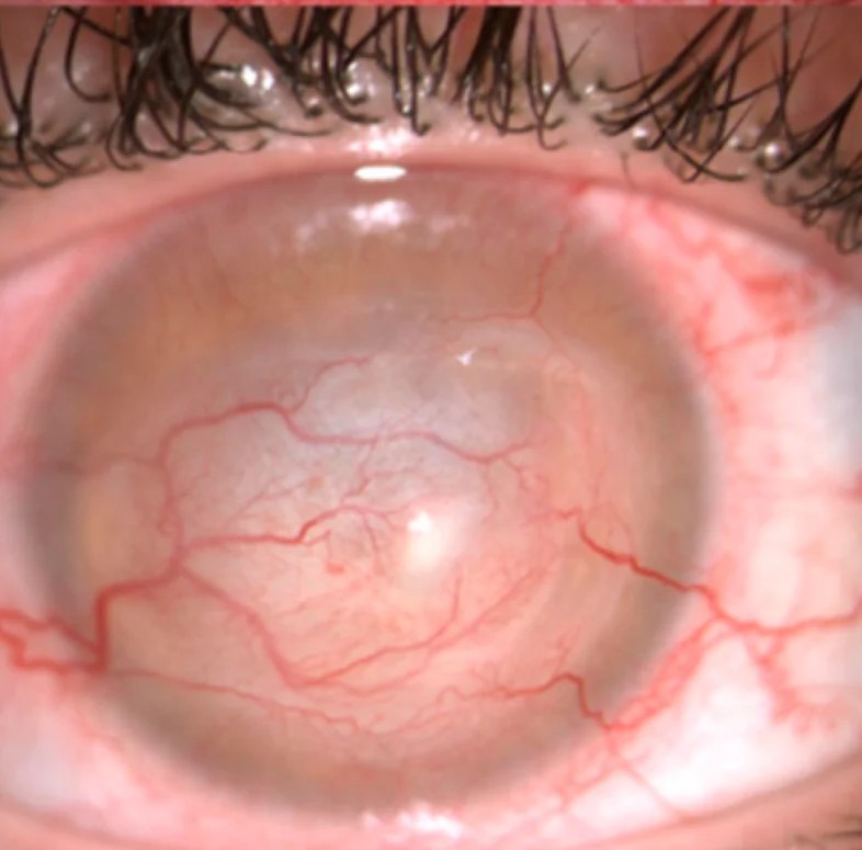
An Acanthamoeba ring infiltrate is a finding no clinician wants to be the first sign that reveals the diagnosis, since its appearance typically marks relatively advanced disease, and the well-documented pattern of delayed or missed diagnosis in Acanthamoeba keratitis, often initially mistaken for herpetic or bacterial keratitis, is precisely why understanding this organism’s early, more subtle signs matters as much as recognizing the ring infiltrate that appears later.
Why This Infection Is So Often Missed Early
Acanthamoeba keratitis frequently begins with symptoms, particularly pain that is often described as disproportionately severe relative to the visible clinical findings, that overlap substantially with more common causes of infectious keratitis, and early examination findings can be nonspecific, sometimes showing only mild epithelial irregularity or a pattern suggestive of herpetic disease, which has historically led to significant, sometimes prolonged, diagnostic delay before the correct cause is identified.
The Ring Infiltrate
As the disease progresses, a ring-shaped stromal infiltrate, reflecting an inflammatory response along the path of radial corneal nerves (radial perineuritis) and the advancing edge of the amoebic infection, becomes a more specific and recognizable sign, though by the time this classic ring appears, the infection has often already caused significant corneal involvement.
- The ring typically represents a zone of host inflammatory response surrounding the advancing organisms, rather than the organisms being confined only to the ring itself
- Radial perineuritis, inflammation tracking along corneal nerve fibers, can produce a characteristic radial or star-like pattern and is considered a relatively specific, though not universally present, sign for this infection
All-fit Slit-Lamp Adapter
Record and share exactly what you see at the slit lamp. One adapter fits any slit lamp or surgical microscope — and any smartphone.
From Choroida — the team behind this siteRisk Factors
- Contact lens wear is the dominant risk factor in most reported series, particularly with exposure to contaminated water, including swimming, showering, or rinsing lenses with tap water while wearing them
- Poor contact lens hygiene more broadly, including use of homemade or improperly prepared saline solutions
- Corneal trauma with exposure to contaminated soil or water in non-contact-lens wearers is a recognized, though less common, alternative risk factor
Earlier, More Subtle Signs
- Disproportionate pain relative to visible clinical findings, a frequently cited early clue
- Epithelial irregularity or a dendritiform pattern that can closely mimic herpetic epithelial keratitis, a common source of early misdiagnosis and inappropriate antiviral-only treatment
- Perineural infiltrates, sometimes visible before a full ring has developed, representing an earlier stage of the same radial perineuritis process
Diagnostic Evaluation
- A specific, deliberate history of contact lens wear and water exposure, or relevant trauma history in non-lens-wearers, given how central risk factor recognition is to raising appropriate suspicion early
- Corneal scraping for culture on specific Acanthamoeba-selective media, and for microscopy, though a negative initial scraping does not exclude the diagnosis given variable organism yield
- In vivo confocal microscopy has become an important tool, allowing direct, non-invasive visualization of Acanthamoeba cysts within the cornea and supporting earlier diagnosis than culture alone in many cases
- Corneal biopsy is considered in cases with strong clinical suspicion but negative scraping and confocal findings, given the serious consequences of a missed or delayed diagnosis
Management
Antiamoebic Therapy
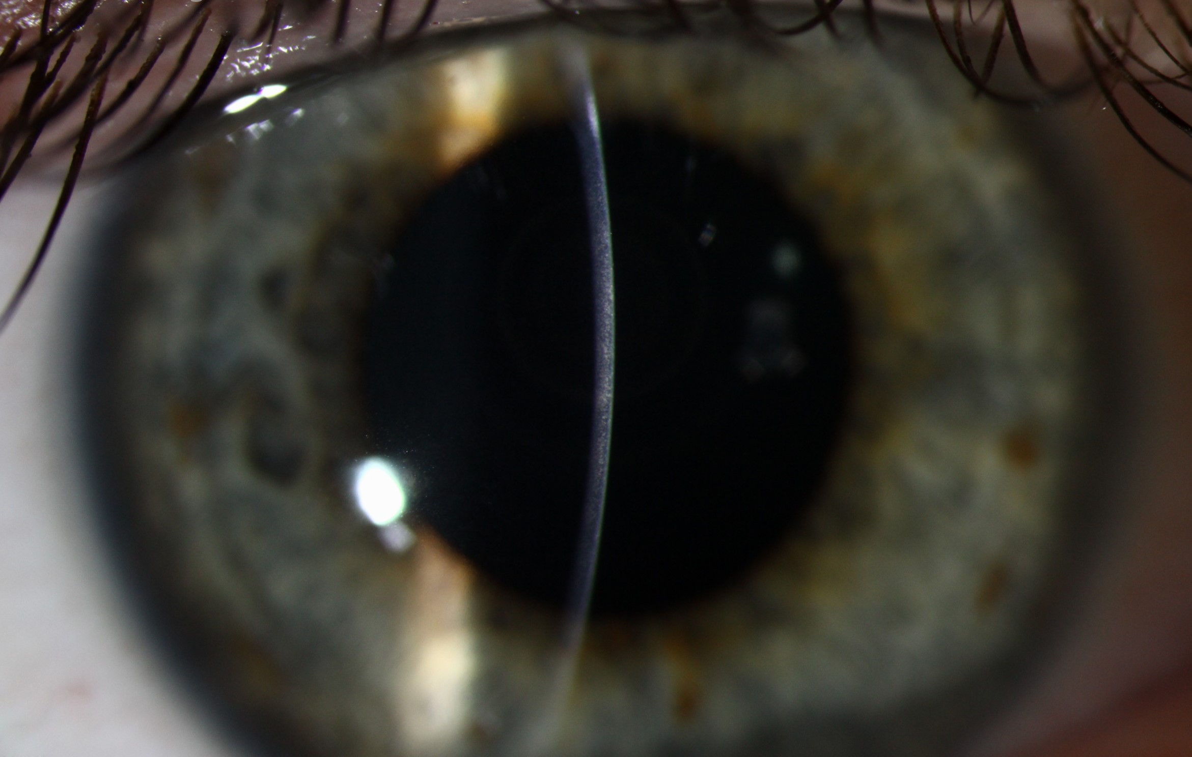
Treatment involves prolonged topical antiamoebic therapy, typically combining a biguanide (such as polyhexamethylene biguanide or chlorhexidine) with a diamidine (such as propamidine), given the resistant cystic form of the organism that makes eradication genuinely difficult and treatment courses correspondingly long, often extending for many months.
Managing Pain and Inflammation
Pain control is an important supportive component given how severe symptoms can be, and the use of topical corticosteroids remains a nuanced, debated area of management, generally considered only after adequate antiamoebic treatment has been established, given concern that premature or unsupported corticosteroid use could worsen the infection.
Surgical Management
Therapeutic keratoplasty is considered for medically refractory disease, corneal perforation, or after infection has been eradicated but left significant visually limiting scarring, though surgery during active infection carries its own specific risks and is generally avoided when medical control can still be achieved.
Why Early Suspicion Matters So Much
Given the well-documented pattern of diagnostic delay in this condition, and given that outcomes correlate strongly with how early appropriate antiamoebic therapy is started relative to disease progression, maintaining a genuinely active index of suspicion, particularly in any contact lens wearer with a keratitis that is not responding as expected to standard antibacterial or antiviral treatment, is one of the single most impactful steps in improving outcomes for this disease.
Prognosis
Early-diagnosed, promptly and adequately treated Acanthamoeba keratitis has a considerably better visual prognosis than disease diagnosed only after significant stromal involvement and ring infiltrate formation have already developed.
This gap in outcomes between early and late diagnosis is the central reason ongoing efforts to raise clinical awareness of this organism’s early, less specific signs, rather than waiting for the more advanced and unmistakable ring infiltrate, remain genuinely important in both clinical practice and ophthalmic education.


Document what you see
Two smartphone imaging tools built for everyday clinic use — one for the slit lamp, one for the fundus.
From Choroida — the team behind this siteReferences
- Dart JK, Saw VP, Kilvington S. Acanthamoeba keratitis: diagnosis and treatment update 2009. Am J Ophthalmol. 2009;148:487-499.
- Lorenzo-Morales J, Khan NA, Walochnik J. An update on Acanthamoeba keratitis: diagnosis, pathogenesis and treatment. Parasite. 2015;22:10.
- Tu EY, Joslin CE, Sugar J, Shoff ME, Booton GC. Prognostic factors affecting visual outcome in Acanthamoeba keratitis. Ophthalmology. 2008;115:1998-2003.
- Kaufman AR, Tu EY. Advances in the management of Acanthamoeba keratitis: a review of the literature and synthesized algorithmic approach. Ocul Surf. 2022;25:26-36.
Test yourself
A few questions straight from this article.
-
What does the appearance of a ring-shaped stromal infiltrate in Acanthamoeba keratitis usually indicate?
The classic ring is a more specific sign, but by the time it appears the infection has often already caused significant corneal involvement, which is why the earlier, subtler signs matter so much. -
What is radial perineuritis in Acanthamoeba keratitis?
Inflammation along the radial corneal nerves produces a radial or star-like pattern and is considered a relatively specific, though not universally present, sign of this infection. -
Which symptom pattern is a frequently cited early clue to Acanthamoeba keratitis?
Pain described as disproportionately severe relative to the visible clinical findings is a frequently cited early clue, at a stage when examination may show only mild epithelial irregularity. -
Which is the dominant risk factor for Acanthamoeba keratitis in most reported series?
Contact lens wear dominates most series, particularly with water exposure such as swimming, showering in lenses, or rinsing lenses with tap water; trauma with contaminated soil or water is a less common alternative. -
Early Acanthamoeba keratitis is most often misdiagnosed as which condition?
Epithelial irregularity or a dendritiform pattern can closely mimic herpetic epithelial keratitis, a common source of early misdiagnosis and inappropriate antiviral-only treatment. -
Which investigation allows direct, non-invasive visualisation of Acanthamoeba cysts within the cornea?
In vivo confocal microscopy visualises cysts directly within the cornea without biopsy and supports earlier diagnosis than culture alone in many cases. -
With strong suspicion of Acanthamoeba keratitis but negative scraping and confocal findings, what is considered next?
Because organism yield from scraping is variable and a negative result does not exclude the diagnosis, corneal biopsy is considered when suspicion stays strong, given the consequences of delay. -
What is the mainstay of medical treatment for Acanthamoeba keratitis?
Prolonged topical therapy typically pairs a biguanide such as polyhexamethylene biguanide or chlorhexidine with a diamidine such as propamidine, often for many months because the cystic form resists eradication. -
When are topical corticosteroids generally considered in Acanthamoeba keratitis?
Corticosteroid use remains a debated area and is generally considered only once adequate antiamoebic treatment is established, because premature or unsupported use could worsen the infection. -
How does the visual prognosis of Acanthamoeba keratitis relate to the timing of diagnosis?
Disease diagnosed and adequately treated early carries a considerably better visual prognosis than disease identified only after significant stromal involvement and ring infiltrate formation.

Writes and reviews the clinical articles on Educate. Every post is checked against peer-reviewed sources before it goes live.


