
Weill-Marchesani syndrome is a rare connective tissue disorder that, in almost every respect, presents as the opposite of Marfan syndrome: short stature instead of tall, brachydactyly instead of arachnodactyly, and a small, round, subluxated lens (microspherophakia) instead of a large, tilted one.
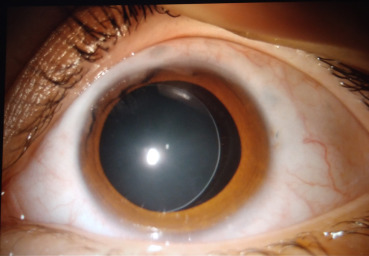
Both autosomal recessive (ADAMTS10, ADAMTS17) and autosomal dominant (FBN1) forms exist, with the gene products involved in extracellular matrix and microfibril regulation, overlapping conceptually with the fibrillin pathway disrupted in Marfan syndrome despite the strikingly different phenotype.
This shared molecular pathway, producing two clinically opposite connective tissue phenotypes, is a useful illustration of how the same broad category of extracellular matrix proteins can manifest in dramatically different ways depending on the specific gene and mutation involved.
Systemic Features
- Short stature, often with a stocky build
- Brachydactyly and joint stiffness, sometimes limiting full extension
- Short, broad hands and feet
- Occasional cardiac involvement (pulmonic stenosis has been reported), though far less prominent than the aortic disease that dominates Marfan syndrome
The body habitus alone is often enough to raise suspicion for Weill-Marchesani syndrome over Marfan syndrome in a patient presenting with lens subluxation, well before any genetic testing is pursued.
This is a genuinely useful bedside distinction, since the two conditions can otherwise share the ectopia lentis finding that first brings a patient to ophthalmic attention, while requiring very different systemic surveillance once the underlying diagnosis is clarified — aortic imaging in Marfan syndrome, joint and cardiac assessment in Weill-Marchesani syndrome.
Ocular Findings
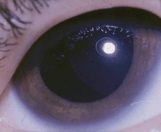
- Microspherophakia — a small, abnormally spherical lens, which itself causes high lenticular myopia even before any subluxation occurs
- Ectopia lentis, in any direction, though inferior displacement is common
- Pupillary block glaucoma — a genuinely dangerous complication, occurring because the small, round, mobile lens can migrate forward and obstruct the pupil far more readily than a normally shaped lens
- High myopia from the abnormal lens shape
- Shallow anterior chamber, compounding the glaucoma risk
The glaucoma risk in Weill-Marchesani syndrome is higher and more acute than in Marfan syndrome, precisely because the spherical shape of the lens allows it to shift forward and block the pupil in a way that a normally shaped, merely displaced lens is less prone to do.
Because acute angle closure from pupillary block can develop with relatively little warning, patients and their families benefit from explicit counseling about the symptoms of an acute attack — sudden eye pain, redness, and blurred vision, sometimes with nausea — so that care is sought urgently rather than after a delay that could risk permanent visual loss.
All-fit Slit-Lamp Adapter
Record and share exactly what you see at the slit lamp. One adapter fits any slit lamp or surgical microscope — and any smartphone.
From Choroida — the team behind this siteDifferential Diagnosis
- Marfan syndrome — tall stature, superotemporal lens subluxation, aortic root disease
- Homocystinuria — marfanoid habitus, inferonasal subluxation, thromboembolic risk
- Isolated microspherophakia — the lens finding without the systemic skeletal features of full Weill-Marchesani syndrome
- Simple ectopia lentis — isolated zonular weakness without an abnormal lens shape
Distinguishing isolated microspherophakia from full Weill-Marchesani syndrome matters practically, since a patient with the lens finding alone still carries the same pupillary block glaucoma risk and needs the same ophthalmic vigilance, even without the short stature and joint findings that would otherwise point toward the complete syndrome.
Diagnostic Evaluation
The diagnosis is largely clinical, based on the combination of short stature, brachydactyly, and the characteristic small, spherical, subluxated lens on slit-lamp exam.
Genetic testing for ADAMTS10, ADAMTS17, or FBN1 mutations can confirm the diagnosis and clarify the inheritance pattern for family counseling, which matters since the recessive and dominant forms carry different recurrence risks.
Gonioscopy and careful assessment of anterior chamber depth are important given the elevated pupillary block risk, and intraocular pressure should be checked and monitored closely, because acute angle closure can develop with less warning than in other causes of ectopia lentis.
A skeletal survey and orthopedic assessment can help characterize the extent of joint involvement once the diagnosis is suspected, and baseline cardiac evaluation is reasonable given the reported, if less prominent, association with pulmonic stenosis.
Management
Miotic agents are used cautiously — and in some cases avoided — because constricting the pupil can actually worsen pupillary block by trapping the spherical lens more securely against the iris.
This is the opposite of how miotics are used in primary angle-closure glaucoma; cycloplegic agents, which pull the lens-iris diaphragm posteriorly, are often preferred instead when the mechanism is lens-related pupillary block.
Laser peripheral iridotomy is used both prophylactically in eyes at high risk and therapeutically for acute pupillary block, creating an alternative pathway for aqueous flow that bypasses the obstructed pupil.
Lens extraction is considered when pupillary block glaucoma is recurrent or difficult to control, or when the lens itself is causing significant visual impairment.
Surgery in these small, unusually shaped, poorly supported lenses requires careful planning given the atypical anatomy, similar in spirit to the technical challenges of any ectopia lentis surgery but complicated further by the spherical shape and elevated angle-closure risk specific to this condition.
Long-term follow-up remains important even after successful treatment of an acute glaucoma episode or lens extraction, since the underlying anatomic predisposition — the abnormal lens shape and shallow anterior chamber — persists and continues to warrant regular intraocular pressure monitoring for the rest of the patient’s life.


Document what you see
Two smartphone imaging tools built for everyday clinic use — one for the slit lamp, one for the fundus.
From Choroida — the team behind this siteReferences
- Faivre L, Gorlin RJ, Wirtz MK, et al. In frame fibrillin-1 gene deletion in autosomal dominant Weill-Marchesani syndrome. Journal of Medical Genetics.
- Khan AO, Aldahmesh MA, Al-Ghadeer H, et al. Familial spherophakia with short stature caused by a novel ADAMTS17 mutation. Ophthalmic Genetics.
- American Academy of Ophthalmology. Basic and Clinical Science Course, Section 11: Lens and Cataract.
- Jones WL. Weill-Marchesani syndrome. Optometry and Vision Science.
Test yourself
A few questions straight from this article.
-
Which body habitus points toward Weill-Marchesani syndrome rather than Marfan syndrome?
Weill-Marchesani syndrome presents as the near-opposite of Marfan syndrome, and the stocky, short-statured habitus with short broad hands is often enough to raise suspicion at the bedside before genetic testing. -
Which genes cause the autosomal recessive form of Weill-Marchesani syndrome?
The recessive form arises from ADAMTS10 or ADAMTS17 mutations, while the dominant form involves FBN1; all three gene products regulate extracellular matrix and microfibrils. -
What refractive consequence does microspherophakia produce even before any lens subluxation?
The abnormally spherical lens shape itself generates high lenticular myopia, so significant refractive error can precede any displacement of the lens. -
Which cardiac lesion has been reported in Weill-Marchesani syndrome?
Occasional cardiac involvement with pulmonic stenosis has been reported, though it is far less prominent than the aortic disease that dominates Marfan syndrome. -
Why is pupillary block glaucoma more acute in Weill-Marchesani syndrome than in Marfan syndrome?
The spherical shape lets the small, mobile lens shift forward and block the pupil far more readily than a normally shaped, merely displaced lens, and a shallow anterior chamber compounds the risk. -
Which topical agents are often preferred for lens-related pupillary block in Weill-Marchesani syndrome?
Cycloplegics pull the lens-iris diaphragm posteriorly, whereas constricting the pupil can worsen block by trapping the spherical lens against the iris; this reverses how miotics are used in primary angle closure. -
How is laser peripheral iridotomy used in Weill-Marchesani syndrome?
Iridotomy creates an alternative route for aqueous that bypasses the obstructed pupil, and it is used both preventively in eyes at high risk and therapeutically during an acute pupillary block attack. -
When is lens extraction considered in Weill-Marchesani syndrome?
Extraction is reserved for recurrent or difficult-to-control pupillary block glaucoma, or when the lens itself causes significant visual impairment; the atypical anatomy makes such surgery demanding. -
What ophthalmic risk does isolated microspherophakia carry without the systemic skeletal features?
A patient with the lens finding alone still carries the same pupillary block glaucoma risk and needs identical ophthalmic vigilance, even without the short stature and joint findings. -
Which features distinguish homocystinuria from Weill-Marchesani syndrome in a patient with ectopia lentis?
Homocystinuria produces a marfanoid build with inferonasal lens subluxation and carries thromboembolic risk, unlike the short, stocky habitus of Weill-Marchesani syndrome.
