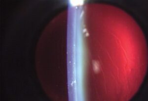
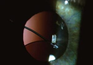
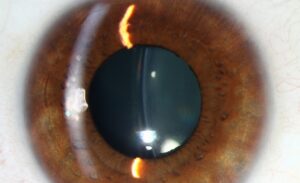
Vitreous loss during cataract surgery, occurring when the posterior capsule ruptures and vitreous gel prolapses forward into the anterior segment or surgical wound, is one of the complications every cataract surgeon trains extensively to recognize and manage, since how promptly and appropriately it is addressed has a direct, measurable impact on the patient’s ultimate visual outcome and risk of further complications.
Why Posterior Capsular Rupture Matters
The posterior capsule normally serves as a barrier separating the anterior segment from the vitreous cavity, and when it ruptures, vitreous gel can prolapse forward, becoming incorporated into the surgical wound, adherent to the iris, or displaced into the anterior chamber.
Unaddressed vitreous in the wound and anterior chamber creates ongoing risks extending well beyond the immediate surgery, including chronic inflammation, cystoid macular edema, pupillary distortion, and, most seriously, an increased long-term risk of retinal detachment from vitreous traction, which is why thorough, meticulous management at the time of the rupture is so important.
Recognizing Vitreous Loss
- A sudden deepening of the anterior chamber or change in the red reflex during phacoemulsification
- Difficulty maintaining chamber stability or an unexpected shift in lens fragment position
- Visible strands of clear, gel-like vitreous, which can be difficult to distinguish visually from aqueous without specific techniques
- A positive finding on gentle sweeping with a cellulose sponge at the wound, where vitreous strands will adhere and can be seen tenting toward the incision, a classic confirmatory sign
Fundus Explorer Pro
Photograph the retinal findings described here with the phone already in your pocket — 22 D optics and built-in illumination in one handheld unit.
From Choroida — the team behind this siteImmediate Intraoperative Management
Stopping and Reassessing
Once vitreous loss is recognized or suspected, the immediate priority is to stop further phacoemulsification or manipulation that could worsen the situation, stabilize the anterior chamber, and reassess before proceeding.
Anterior Vitrectomy
Thorough removal of vitreous from the anterior chamber and wound, using a vitrectomy handpiece with appropriate cut and low-flow settings, is the central step in management, aiming to clear all vitreous from the anterior segment and wound to prevent the complications associated with retained vitreous.
- A dispersive ophthalmic viscosurgical device is often used to help stabilize the anterior chamber and push vitreous posteriorly, supporting safe vitrectomy
- Triamcinolone can be used intraoperatively to stain and visualize otherwise transparent vitreous strands, substantially improving the surgeon’s ability to confirm complete removal
Managing Retained Lens Material
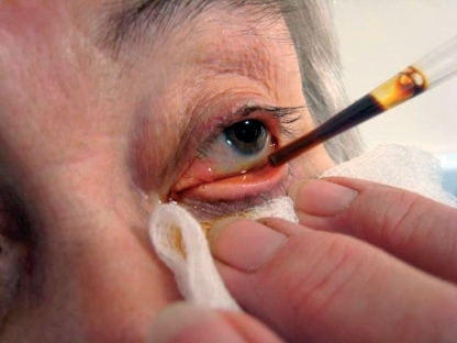
Any remaining lens fragments must be removed carefully, avoiding pushing fragments posteriorly into the vitreous cavity, since a retained lens fragment in the posterior segment generally requires a separate vitreoretinal procedure for removal and can cause significant postoperative inflammation if left unaddressed.
Intraocular Lens Placement Decisions
The presence and extent of posterior capsular support remaining after the rupture and vitrectomy determines the safest intraocular lens fixation strategy.
- If adequate capsular support remains, in-the-bag or sulcus placement of the intraocular lens can often still be achieved
- With more extensive capsular loss, sulcus fixation with optic capture through an intact anterior capsulorhexis, when feasible, provides good stability
- In eyes with insufficient capsular support for either of the above, alternative fixation strategies, including a scleral-fixated or, in select cases, an anterior chamber intraocular lens, are considered
Postoperative Management
Eyes with vitreous loss require closer postoperative monitoring than routine cataract surgery, given the increased risk of cystoid macular edema, elevated intraocular pressure, and, over the longer term, retinal detachment, and patients are counseled specifically about the importance of prompt reporting of new floaters, flashes, or a visual field defect suggestive of retinal detachment.
Prognosis
With prompt recognition and thorough, meticulous management, including complete vitrectomy and appropriate intraocular lens fixation planning, most eyes with vitreous loss during cataract surgery achieve good visual outcomes, though the complication does carry a real, measurably increased long-term risk of retinal detachment and other complications compared with uncomplicated cataract surgery, supporting the closer follow-up these patients receive.


Document what you see
Two smartphone imaging tools built for everyday clinic use — one for the slit lamp, one for the fundus.
From Choroida — the team behind this siteReferences
- Osher RH, Cionni RJ. The torn posterior capsule: its intraoperative behavior, surgical management, and long-term consequences. J Cataract Refract Surg. 1990;16:490-494.
- Vajpayee RB, Sharma N, Dada T, Gupta V, Kumar A, Dada VK. Management of posterior capsule tears. Surv Ophthalmol. 2001;45:473-488.
- Ionides A, Minassian D, Tuft S. Visual outcome following posterior capsule rupture during cataract surgery. Br J Ophthalmol. 2001;85:222-224.
- Wong TY, Foster PJ, Johnson GJ, Seah SK. Risk factors for and outcomes of cataract surgery complicated by posterior vitreous loss. Am J Ophthalmol. 2003;136:296-305.
Test yourself
A few questions straight from this article.
-
What role does an intact posterior capsule normally serve during cataract surgery?
The posterior capsule normally separates the anterior segment from the vitreous cavity; once it ruptures, vitreous gel can prolapse forward into the chamber or wound. -
Which manoeuvre gives the classic confirmatory sign of vitreous at the cataract wound?
Vitreous strands adhere to a cellulose sponge swept gently at the wound and can be seen tenting toward the incision, the classic confirmatory sign. -
Which change during phacoemulsification should raise suspicion of posterior capsular rupture?
A sudden deepening of the chamber or an altered red reflex, together with difficulty keeping the chamber stable, are early intraoperative cues to a capsular break. -
What is the most serious long-term risk of vitreous left unaddressed in the wound and anterior chamber?
Retained vitreous in the wound exerts traction and carries an increased long-term risk of retinal detachment, the most serious of several consequences that also include chronic inflammation, cystoid macular oedema and pupillary distortion. -
Once vitreous loss is recognised during cataract surgery, what is the immediate priority?
The first step is to stop any manipulation that could worsen the break, stabilise the anterior chamber, and reassess before continuing. -
Which agent is used intraoperatively to stain and visualise transparent vitreous strands?
Triamcinolone stains otherwise transparent vitreous, substantially improving the surgeon's ability to confirm that removal is complete. -
Why is a dispersive viscosurgical device used during management of vitreous loss?
A dispersive ophthalmic viscosurgical device helps hold the anterior chamber and displaces vitreous posteriorly, supporting a safe anterior vitrectomy. -
What is the consequence of a lens fragment being pushed posteriorly into the vitreous cavity?
A retained fragment in the posterior segment usually needs a separate vitreoretinal operation and can cause significant postoperative inflammation if left in place. -
With extensive capsular loss but an intact anterior capsulorhexis, which lens fixation gives good stability?
When capsular loss is more extensive, sulcus placement with optic capture through an intact anterior capsulorhexis provides good stability where it is feasible. -
After cataract surgery complicated by vitreous loss, which symptoms are patients told to report promptly?
These eyes carry an increased long-term risk of retinal detachment, so patients are counselled specifically to report new floaters, flashes or a field defect without delay.

Writes and reviews the clinical articles on Educate. Every post is checked against peer-reviewed sources before it goes live.