
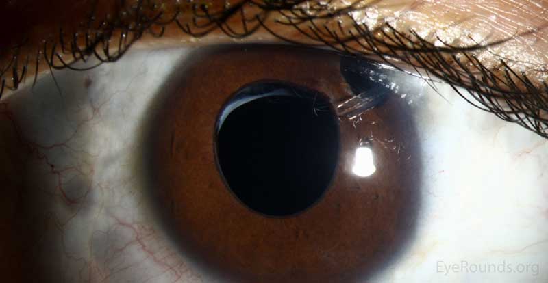
Aphakic glaucoma is elevated intraocular pressure developing in an eye that has had its natural lens removed without an intraocular lens implant, most classically after pediatric cataract surgery performed in infancy or early childhood.
It is distinct in both mechanism and long-term behavior from the more familiar pseudophakic and general secondary glaucomas seen after routine adult cataract surgery, and it carries its own specific, lifelong surveillance requirements.
Because pediatric aphakic glaucoma can develop silently over years and its presentation differs meaningfully from adult glaucoma, lifelong monitoring rather than a fixed postoperative follow-up window is the standard of care.
It affects a meaningful proportion of children who undergo lensectomy without an implant, making it one of the more consequential long-term risks families need to understand at the time of the original cataract surgery, not just an abstract future possibility.
Why Pediatric Aphakia Differs
Aphakic glaucoma is overwhelmingly a pediatric phenomenon, since modern adult cataract surgery routinely includes intraocular lens implantation, while a meaningful proportion of pediatric cataract surgery, particularly in very young infants, is still performed without a primary lens implant due to the challenges of accurate power calculation in a rapidly growing eye.
The risk is highest when cataract surgery is performed very early in infancy, and it persists for years to decades after the original surgery rather than being confined to an early postoperative window, distinguishing it sharply from most other postsurgical glaucomas.
This prolonged, unpredictable time course is the central reason aphakic glaucoma requires a fundamentally different monitoring strategy than adult postoperative glaucoma.
Mechanism
The exact mechanism remains incompletely understood, but proposed contributors include abnormal angle development altered by the absence of the lens, chronic low-grade inflammation from the original surgery, and mechanical changes to the trabecular meshwork related to the aphakic state itself.
Vitreous or other tissue can also mechanically affect angle structures in some aphakic eyes, contributing to outflow obstruction through a more direct structural pathway.
Because multiple mechanisms likely contribute in different proportions across different patients, no single unifying explanation accounts for every case, which is part of why the condition remains difficult to predict reliably for any individual child.
This unpredictability is exactly why surveillance cannot be safely limited to children thought to be at highest risk, since even eyes without obvious additional risk factors have gone on to develop significant pressure elevation years later.
All-fit Slit-Lamp Adapter
Record and share exactly what you see at the slit lamp. One adapter fits any slit lamp or surgical microscope — and any smartphone.
From Choroida — the team behind this siteRisk Factors
- Surgery performed in early infancy, particularly within the first few months of life, the single strongest known risk factor
- Microcornea or other associated anterior segment anomalies present at the time of the original cataract
- A longer time since surgery, since risk accumulates over years rather than being confined to the early postoperative period
- Additional intraocular surgery or complications during the original cataract procedure
Clinical Presentation and Detection
Aphakic glaucoma is frequently asymptomatic, especially in a young child who cannot reliably report symptoms, making scheduled surveillance rather than symptom-triggered evaluation essential to catching it early.
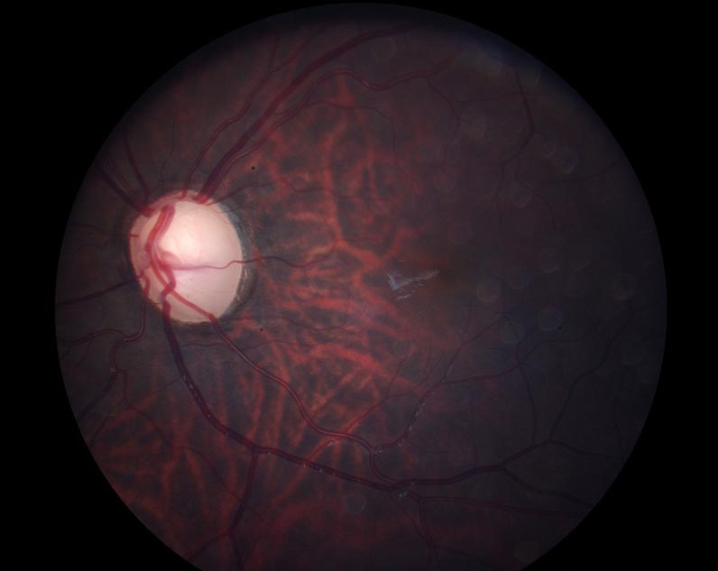
Signs to watch for include an enlarging corneal diameter or axial length in a very young child whose eye can still expand under elevated pressure, progressive myopic shift, optic disc changes, and, in an older child, visual field loss detectable on formal testing.
Because pressure elevation can develop many years after the original surgery, and because a young child’s eye can still enlarge in response to elevated pressure in a way an adult eye cannot, buphthalmos-like globe enlargement remains a relevant clinical sign well beyond early infancy in these patients.
Management
Lifelong, regular monitoring of intraocular pressure, corneal diameter or axial length in young children, and optic nerve status is the cornerstone of managing any eye that has undergone lensectomy without implant, continuing indefinitely rather than tapering off after a fixed postoperative period.
Topical glaucoma medications are used first-line when elevated pressure is detected, following similar principles to adult glaucoma management but requiring particular attention to pediatric dosing and side-effect considerations.
Surgical intervention, including angle surgery, trabeculectomy, or a glaucoma drainage device, discussed in its own dedicated article on this site, is used for eyes that fail adequate medical control, following a similar general treatment ladder to other forms of pediatric glaucoma.
Coordinating this lifelong surveillance across a family’s changing pediatric and then adult ophthalmology care, often spanning multiple providers over decades, deserves deliberate attention, since gaps in follow-up are exactly when a slowly progressive, asymptomatic glaucoma is most likely to go unnoticed.


Document what you see
Two smartphone imaging tools built for everyday clinic use — one for the slit lamp, one for the fundus.
From Choroida — the team behind this siteReferences
- Infant Aphakia Treatment Study Group. Glaucoma-related adverse events in the first 5 years after unilateral cataract removal in the Infant Aphakia Treatment Study. JAMA Ophthalmology.
- American Academy of Ophthalmology. Basic and Clinical Science Course, Section 6: Pediatric Ophthalmology and Strabismus.
- Chen TC, Bhatia LS, Halpern EF, Walton DS. Risk factors for the development of aphakic glaucoma after congenital cataract surgery. Transactions of the American Ophthalmological Society.
Test yourself
A few questions straight from this article.
-
Aphakic glaucoma develops in eyes with which surgical history?
Aphakic glaucoma is raised intraocular pressure in an eye whose natural lens has been removed without an intraocular lens implant, most classically after pediatric cataract surgery. -
Why is aphakic glaucoma overwhelmingly a pediatric rather than an adult problem?
Modern adult surgery routinely implants a lens, while much pediatric surgery in very young infants is still done without a primary implant because power calculation in a rapidly growing eye is difficult. -
Which factor is the single strongest known risk for developing aphakic glaucoma?
Surgery in early infancy, particularly within the first few months of life, is described as the single strongest known risk factor for later aphakic glaucoma. -
Which is a proposed contributing mechanism in aphakic glaucoma?
Proposed contributors include abnormal angle development related to the missing lens, chronic low-grade postoperative inflammation, and mechanical changes to the trabecular meshwork. -
Why is surveillance for aphakic glaucoma not restricted to children judged highest risk?
The condition is unpredictable for any individual child, and eyes without obvious additional risk factors have gone on to develop significant pressure elevation years after surgery. -
Which finding suggests aphakic glaucoma in a very young child after lensectomy?
Because the condition is usually asymptomatic in a young child, monitored signs include enlarging corneal diameter or axial length, progressive myopic shift and optic disc changes. -
Why does globe enlargement remain a useful sign well beyond early infancy in these patients?
A young child's eye can still enlarge in response to raised pressure in a way an adult eye cannot, so buphthalmos-like enlargement stays clinically relevant beyond early infancy. -
What is the first-line treatment when raised pressure is detected in an aphakic child?
Topical glaucoma medications are used first-line, following adult principles but with particular attention to pediatric dosing and side-effect considerations. -
What is the next step for an aphakic eye that fails adequate medical pressure control?
Eyes failing medical control move to surgery, including angle surgery, trabeculectomy or a drainage device, following the general treatment ladder used in other pediatric glaucomas. -
What defines the correct follow-up strategy after pediatric lensectomy without a lens implant?
Because risk persists for years to decades and the condition is silent, lifelong monitoring of intraocular pressure, corneal diameter or axial length, and optic nerve status continues indefinitely.
