
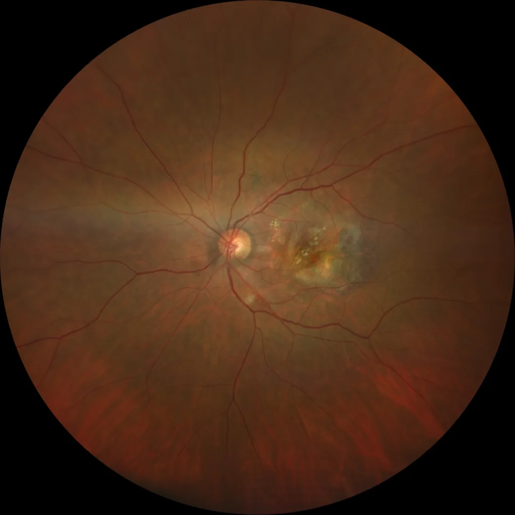
Retinal pigment epithelial (RPE) tear is a recognized complication of neovascular age-related macular degeneration, occurring most often in eyes with a large pigment epithelial detachment (PED) that is being treated with anti-VEGF therapy, and it represents a genuine and somewhat frustrating clinical paradox — the very treatment that successfully controls choroidal neovascularization and reduces fluid can, in certain higher-risk eyes, precipitate a sudden, often vision-limiting tear of the RPE layer itself.
Mechanism
A large fibrovascular or serous PED creates significant tangential (shearing) stress across the RPE monolayer as it is stretched over an elevated dome of subretinal fluid or fibrovascular tissue.
Anti-VEGF treatment rapidly reduces fluid and induces contraction of the underlying choroidal neovascular membrane, and this sudden contraction can generate enough additional shearing force across an already stretched, structurally vulnerable RPE layer to cause it to tear — typically at the junction between the still-attached and detached RPE, where the mechanical stress is concentrated.
The RPE layer then retracts, exposing bare choroid in the area of the tear while the retracted RPE tissue rolls up elsewhere, usually still attached at one edge.
Risk Factors
- Large PED height and diameter — the single most consistently identified risk factor, with larger, more elevated PEDs carrying substantially higher tear risk than smaller ones
- A predominantly serous (rather than purely fibrovascular) PED composition
- The specific type and characteristics of the underlying choroidal neovascular membrane
- Rapid, robust treatment response to anti-VEGF therapy — somewhat counterintuitively, eyes that respond most dramatically to treatment, with rapid PED collapse, appear to carry a higher tear risk than those with a more gradual response
Given this risk profile, patients with a large PED being started on anti-VEGF therapy should be counseled about the possibility of RPE tear as a recognized complication before treatment begins, so that any sudden change in vision after an injection is reported and evaluated promptly rather than assumed to simply reflect expected treatment variability.
Fundus Explorer Pro
Photograph the retinal findings described here with the phone already in your pocket — 22 D optics and built-in illumination in one handheld unit.
From Choroida — the team behind this siteClinical Presentation
Patients typically notice sudden, sometimes significant, painless vision loss, often within days of an anti-VEGF injection, corresponding to the timing of PED contraction described above.
Because the underlying disease (neovascular AMD) already causes some baseline visual impairment, a new, more abrupt change should be specifically recognized as potentially representing a tear rather than simply attributed to disease progression or normal fluctuation.
Exam and Imaging Findings
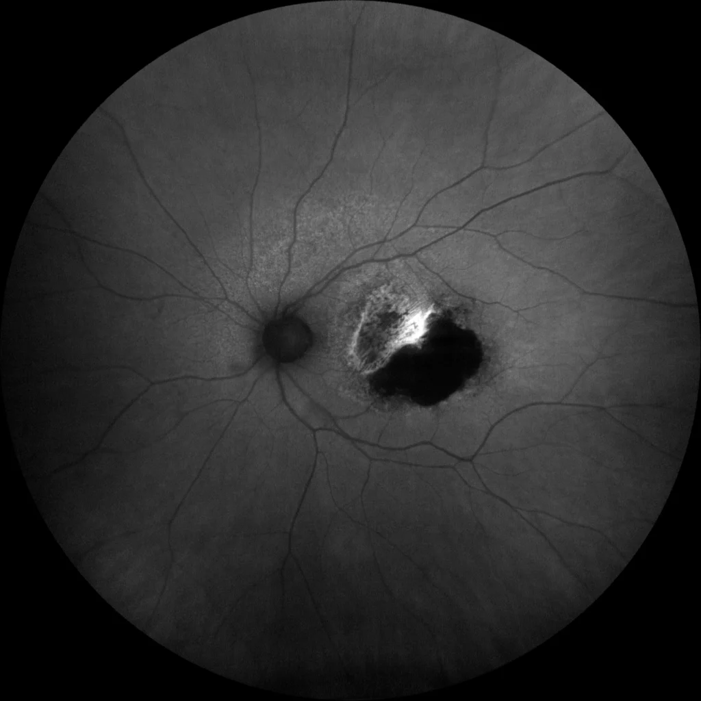
- A sharply demarcated area of exposed, depigmented choroid where the RPE has torn away, often with a visible, rolled or scrolled edge of retracted RPE tissue at the margin, sometimes appearing as a crescentic or horseshoe-shaped darker band adjacent to the pale, exposed area
- OCT clearly demonstrates the RPE discontinuity, showing the retracted, folded RPE layer and the adjacent area where the RPE-Bruch’s membrane complex is now absent, with the underlying choroid directly visible
- The area of RPE tear, since the RPE normally forms part of the outer blood-retina barrier and also nourishes the overlying photoreceptors, generally corresponds to permanently reduced function if the tear involves the fovea, regardless of how well the underlying neovascular activity is subsequently controlled
Differential Diagnosis
- Progression of the underlying choroidal neovascularization or geographic atrophy — a more gradual process than the typically sudden onset of a true RPE tear, and without the specific rolled-edge, exposed-choroid appearance characteristic of a tear on exam and OCT
- Submacular hemorrhage — a different, though sometimes coexisting, complication of neovascular AMD, distinguished by the presence of blood rather than a clean area of RPE discontinuity
- Central serous chorioretinopathy-related PED changes — a different underlying disease process with its own characteristic findings, discussed in this site’s coverage of that condition
Management
There is no treatment that repairs or reverses an established RPE tear itself; management focuses on continuing appropriate anti-VEGF therapy for any persistent or recurrent choroidal neovascular activity in the areas surrounding the tear, since the underlying neovascular disease still requires ongoing control even though the tear itself cannot be undone.
Some clinicians consider adjusting anti-VEGF treatment strategy (such as considering a somewhat more gradual treatment approach or closer monitoring) in high-risk eyes with a large PED, given the recognized association between rapid treatment response and tear risk, though this needs to be balanced against the clear overall benefit of anti-VEGF treatment for controlling the neovascular disease itself.
Prognosis
Visual outcome after RPE tear depends heavily on whether the fovea is involved in the torn (rather than the retracted, still-present) area — tears sparing central foveal RPE can be associated with relatively preserved central vision, while foveal involvement generally results in permanent, significant central visual impairment given the RPE’s essential supportive role for the overlying photoreceptors.
Because of this direct relationship between tear location and visual outcome, and because tears can occur even with appropriate, guideline-concordant anti-VEGF treatment, patient counseling about this risk — rather than any specific preventive strategy with strong proven efficacy — remains the primary practical tool available before treatment begins in a high-risk eye, so that the possibility is understood in advance rather than discovered only as an unexpected, alarming complication.


Document what you see
Two smartphone imaging tools built for everyday clinic use — one for the slit lamp, one for the fundus.
From Choroida — the team behind this siteReferences
- Chan CK, Abraham P, Meyer CH, et al. Optical coherence tomography-measured pigment epithelial detachment height as a predictor for retinal pigment epithelial tears associated with intravitreal bevacizumab injections. Retina.
- Clemens CR, Bastian N, Alten F, et al. Prediction of retinal pigment epithelial tear in serous pigment epithelial detachments. Acta Ophthalmologica.
- American Academy of Ophthalmology. Basic and Clinical Science Course, Section 12: Retina and Vitreous.
Test yourself
A few questions straight from this article.
-
In which clinical setting does a retinal pigment epithelial tear most often occur?
RPE tear is a recognised complication of neovascular AMD, occurring most often in eyes with a large pigment epithelial detachment being treated with anti-VEGF therapy. -
How does anti-VEGF therapy precipitate a retinal pigment epithelial tear?
Anti-VEGF rapidly reduces fluid and contracts the choroidal neovascular membrane, generating extra shearing force across an RPE monolayer already stretched over the elevated PED. -
Where does a retinal pigment epithelial tear typically occur within the detachment?
The tear typically occurs at the junction between the still-attached and the detached RPE, where the mechanical shearing stress is concentrated. -
Which feature of a pigment epithelial detachment is the most consistently identified risk factor for tear?
Large PED height and diameter is the single most consistently identified risk factor, with larger, more elevated detachments carrying substantially higher tear risk. -
Which treatment response pattern is counterintuitively associated with a higher RPE tear risk?
Eyes that respond most dramatically to anti-VEGF, with rapid PED collapse, appear to carry a higher tear risk than those whose response is more gradual. -
How does a retinal pigment epithelial tear typically present?
Patients typically notice sudden, sometimes marked, painless vision loss within days of an anti-VEGF injection, matching the timing of PED contraction. -
Which fundus appearance indicates a retinal pigment epithelial tear?
A tear shows a sharply demarcated area of exposed, depigmented choroid, with a visible rolled or scrolled edge of retracted RPE forming a darker crescentic band at the margin. -
Which feature separates submacular haemorrhage from a retinal pigment epithelial tear?
Submacular haemorrhage is a different, sometimes coexisting complication of neovascular AMD, identified by blood rather than by a clean area of RPE discontinuity. -
What most strongly determines the visual outcome after a retinal pigment epithelial tear?
Tears sparing the central foveal RPE can leave central vision relatively preserved, while foveal involvement generally causes permanent, significant central impairment. -
How is an established retinal pigment epithelial tear managed?
Nothing repairs the tear itself, so management continues appropriate anti-VEGF therapy for persistent or recurrent neovascular activity around the tear, since the underlying disease still needs control.

Writes and reviews the clinical articles on Educate. Every post is checked against peer-reviewed sources before it goes live.


