
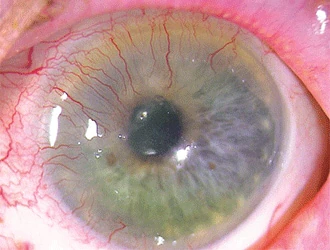
Corneal neovascularization is the growth of new blood vessels into the normally avascular cornea, a change that is never itself a diagnosis but is always a sign of some underlying disruption to the cornea’s carefully maintained avascular state.
The cornea’s clarity depends on remaining free of blood vessels, and any process that tips the balance of angiogenic and anti-angiogenic factors toward new vessel growth threatens that clarity directly, which is why neovascularization deserves attention regardless of how mild it initially appears.
Identifying the underlying cause, rather than treating the vessels themselves as the primary problem, is the essential first step whenever corneal neovascularization is found on exam.
Even mild, peripheral vascularization is worth documenting and monitoring over time, since early, subtle changes are far easier to reverse or stabilize than established, deep vessels found only once they have already become visually significant.
Why the Cornea Is Normally Avascular
The cornea maintains an active balance of pro-angiogenic and anti-angiogenic factors that normally favors avascularity, a state essential for optical clarity since blood vessels and the fluid leakage or scarring associated with them would otherwise scatter and block light.
Chronic hypoxia, inflammation, or limbal stem cell deficiency can tip this balance, allowing vessels to sprout from the limbal vascular arcade and grow inward across what is normally clear, avascular tissue.
Once vessels are established, they rarely regress completely spontaneously, even after the inciting cause is addressed, which is part of why prevention and early intervention are generally more effective than waiting to treat established neovascularization.
Lymphatic vessels frequently accompany the blood vessels in this process, a less visible but clinically important component that contributes independently to the elevated graft rejection risk discussed further below.
Causes
- Chronic contact lens wear, particularly extended or overnight wear, causing relative corneal hypoxia that is one of the most common causes encountered in routine practice
- Chronic ocular surface inflammation, including severe or longstanding blepharitis, vernal keratoconjunctivitis, or atopic keratoconjunctivitis
- Infectious keratitis, particularly herpetic disease, discussed in relation to herpes simplex keratitis in its own dedicated article on this site, where inflammation and tissue damage can drive vessel ingrowth
- Limbal stem cell deficiency, from any cause, disrupting the normal limbal barrier that helps maintain corneal avascularity
- Chemical injury or severe corneal thinning disorders, where tissue damage provokes a reparative but ultimately clarity-threatening vascular response
All-fit Slit-Lamp Adapter
Record and share exactly what you see at the slit lamp. One adapter fits any slit lamp or surgical microscope — and any smartphone.
From Choroida — the team behind this siteClinical Patterns
Superficial neovascularization involves vessels growing within the epithelium and anterior stroma, most classically from chronic contact lens-related hypoxia, and tends to be more responsive to addressing the underlying cause.
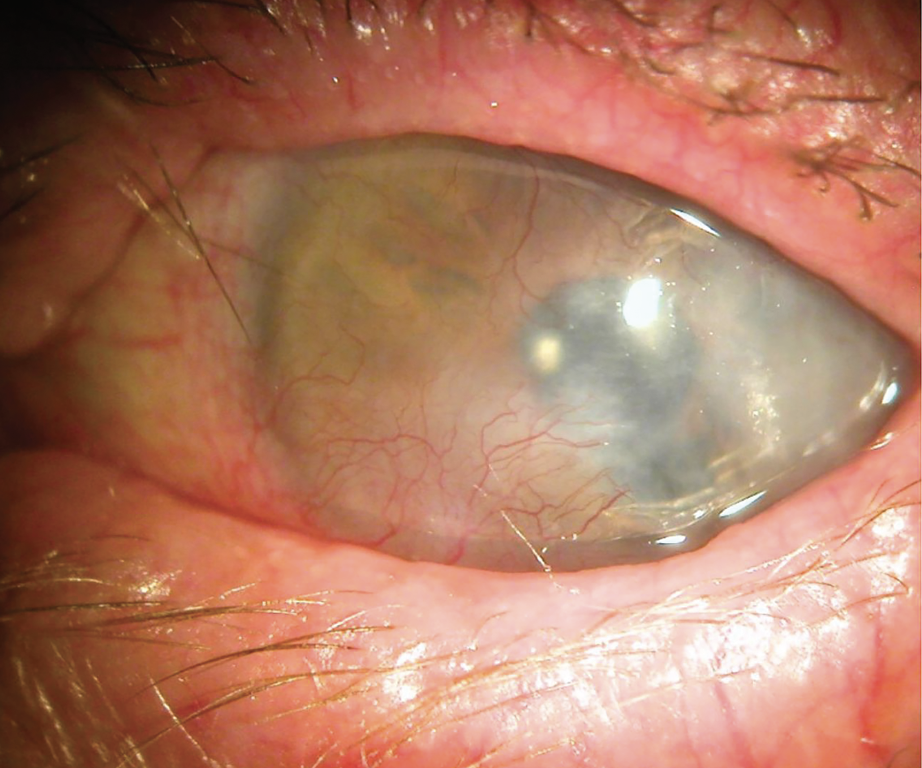
Deep, or stromal, neovascularization involves vessels extending into the deeper corneal stroma, typically from a more severe inflammatory or infectious process, and tends to be more persistent and more visually significant given its proximity to the visual axis in central cases.
Ghost vessels, non-perfused but structurally intact vessel remnants visible after an episode of active neovascularization has resolved, remain relevant clinically since they can rapidly reperfuse if the same inciting stimulus recurs.
Why It Matters for Vision
New vessels themselves can directly obscure vision when they extend into the central visual axis, and the associated lipid exudation, edema, and scarring that often accompany them compound this direct visual impact.
Corneal neovascularization also carries indirect risk for future surgery: a vascularized cornea has a substantially higher risk of graft rejection after penetrating keratoplasty or endothelial keratoplasty, both discussed in their own dedicated articles on this site, since the new vessels provide direct immune cell access to donor tissue that an avascular cornea normally lacks.
This elevated rejection risk is a major reason ophthalmologists try to minimize or eliminate corneal vascularization before elective corneal transplant surgery whenever that is achievable.
Management
Addressing the underlying cause is the essential first step: switching contact lens modality or discontinuing extended wear, controlling chronic ocular surface inflammation, or treating an active infectious process.
Topical corticosteroids can reduce active inflammatory neovascularization, particularly useful for recently established, still-active vessels rather than long-standing, mature ones.
Anti-VEGF therapy, delivered topically or by subconjunctival injection, has shown benefit in some cases of active corneal neovascularization, targeting the angiogenic signaling pathway directly, though its use here remains somewhat more specialized and less standardized than its established role in retinal disease.
Fine needle diathermy or laser photocoagulation of established feeder vessels is used selectively for mature, well-defined neovascularization that has not responded adequately to medical therapy, particularly when reducing vascularization before a planned corneal transplant.
Regardless of which treatment is chosen, ongoing surveillance remains important, since the underlying angiogenic drive can persist even after visible vessels have been treated, and recurrence is a genuine possibility if the original inciting factor is not fully controlled.


Document what you see
Two smartphone imaging tools built for everyday clinic use — one for the slit lamp, one for the fundus.
From Choroida — the team behind this siteReferences
- Chang JH, Gabison EE, Kato T, Azar DT. Corneal neovascularization. Current Opinion in Ophthalmology.
- American Academy of Ophthalmology. Basic and Clinical Science Course, Section 8: External Disease and Cornea.
- Cursiefen C, Chen L, Dana MR, Streilein JW. Corneal lymphangiogenesis: evidence, mechanisms, and implications for corneal transplant immunology. Cornea.
Test yourself
A few questions straight from this article.
-
How should corneal neovascularization be interpreted when found on slit lamp examination?
Corneal neovascularization is never itself a diagnosis. It always signals some underlying process that has disrupted the cornea's normally avascular state, so identifying that cause is the first step. -
From which structure do new vessels sprout when the cornea loses its avascular state?
When chronic hypoxia, inflammation or limbal stem cell deficiency tips the angiogenic balance, vessels sprout from the limbal vascular arcade and grow inward across normally clear tissue. -
Which cause of corneal neovascularization is among the most common encountered in routine practice?
Chronic contact lens wear, particularly extended or overnight wear, produces relative corneal hypoxia and is one of the most common causes of corneal neovascularization seen in routine practice. -
Superficial corneal neovascularization involves vessels growing within which corneal layers?
Superficial neovascularization runs within the epithelium and anterior stroma, most classically from chronic contact lens-related hypoxia, and it tends to respond better once the underlying cause is addressed. -
What are ghost vessels in a cornea previously affected by neovascularization?
Ghost vessels are non-perfused yet structurally intact remnants visible after active neovascularization resolves. They matter because they can rapidly reperfuse if the same inciting stimulus recurs. -
Why does a vascularized cornea carry a higher risk of graft rejection after keratoplasty?
A vascularized cornea has a substantially higher rejection risk after penetrating or endothelial keratoplasty because the new vessels provide immune cells direct access to donor tissue that an avascular cornea lacks. -
What is the essential first step in managing corneal neovascularization?
Management begins with the cause: changing contact lens modality or stopping extended wear, controlling chronic ocular surface inflammation, or treating an active infection. -
Which treatment is used selectively for mature, well-defined corneal vessels unresponsive to medical therapy?
Fine needle diathermy or laser photocoagulation of feeder vessels is reserved for mature, well-defined neovascularization that has not responded to medical therapy, particularly before a planned corneal transplant. -
Besides blood vessels, which accompanying vessels contribute to graft rejection risk in corneal neovascularization?
Lymphatic vessels frequently accompany the new blood vessels. They are less visible on examination but contribute independently to the elevated graft rejection risk. -
What is the expected behaviour of established corneal vessels once the inciting cause is treated?
Established corneal vessels seldom regress fully by themselves even after the inciting cause is addressed, which is why prevention and early intervention outperform treating mature neovascularization.

Writes and reviews the clinical articles on Educate. Every post is checked against peer-reviewed sources before it goes live.


