
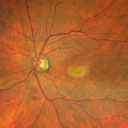
Adult-onset vitelliform maculopathy is a distinct entity from Best vitelliform macular dystrophy, sharing a similar egg-yolk-like appearance on fundus exam but differing in nearly every other respect that matters clinically: later age of onset, generally milder and slower visual impact, a smaller lesion size, and, critically, a normal or only mildly abnormal electro-oculogram, in sharp contrast to the severely abnormal EOG that is a defining diagnostic feature of true Best disease.
Confusing the two conditions leads to unnecessary alarm about progression and inheritance that adult-onset disease does not carry to the same degree, which is why getting the distinction right at the time of diagnosis matters as much for patient counseling as it does for clinical management.
Why the Distinction From Best Disease Matters
Best vitelliform macular dystrophy is a childhood-onset, BEST1-related inherited condition with a severely abnormal EOG (reflecting widespread retinal pigment epithelial dysfunction well beyond the visible macular lesion) and a generally more progressive course through recognized clinical stages toward eventual macular atrophy or choroidal neovascularization.
Adult-onset vitelliform maculopathy, presenting instead in middle age or later, typically shows a normal or only mildly subnormal EOG, a smaller and often more centrally located lesion, and a course that, while not entirely benign, generally progresses more slowly and with less severe eventual visual impact than classic Best disease.
Pathogenesis
The exact mechanism remains less clearly defined than in Best disease, though it is thought to relate to focal dysfunction of the retinal pigment epithelium leading to accumulation of lipofuscin-like material beneath the fovea, producing the characteristic round, yellow, subretinal deposit.
Some cases have been associated with mutations in genes including PRPH2 or, in a smaller subset, BEST1 itself, suggesting the condition may represent a genetically and mechanistically heterogeneous group rather than a single uniform disease entity.
However, the majority of cases remain without a clearly identified genetic cause on routine testing.
This genetic heterogeneity, combined with the predominantly sporadic pattern seen in most patients, is part of why adult-onset vitelliform maculopathy is generally regarded as a distinct clinical entity or final common pathway rather than a single, well-defined single-gene disorder in the way Best disease is.
Fundus Explorer Pro
Photograph the retinal findings described here with the phone already in your pocket — 22 D optics and built-in illumination in one handheld unit.
From Choroida — the team behind this siteClinical Presentation
Patients typically present in their 40s to 60s with mild, gradual central vision blurring or metamorphopsia, often first noticed on an Amsler grid or during a routine eye exam rather than as a dramatic, acute visual complaint.
Vision is frequently preserved at a reasonably good level for years, and many patients remain only mildly symptomatic despite the visible macular finding.
This can itself be reassuring information to relay once the diagnosis is established, particularly for a patient who may have searched online and encountered alarming information about Best disease before understanding that their own condition follows a different, generally milder course.
Exam and Imaging Findings
- A round or oval, yellow, subfoveal or juxtafoveal lesion, typically smaller than the classic egg-yolk lesion of childhood Best disease
- OCT showing a dome-shaped subretinal deposit between the retinal pigment epithelium and photoreceptors, corresponding to the visible yellow material on fundus exam
- Normal or only mildly reduced EOG light rise, distinguishing it from the severely abnormal EOG of true Best disease
- Fundus autofluorescence showing hyperautofluorescence of the lesion, from accumulated lipofuscin-like material, useful both for diagnosis and for monitoring change over time
- Absence of the multifocal or widespread pattern sometimes seen in some Best disease variants
Differential Diagnosis
- Best vitelliform macular dystrophy — childhood onset, severely abnormal EOG, typically a family history and staged progression, discussed in more detail in this site’s dedicated coverage of Best disease
- Pattern dystrophy of the retinal pigment epithelium — a related but distinct group of conditions with their own characteristic appearances, some of which can show overlapping features
- Age-related macular degeneration with a pseudovitelliform pigment epithelial detachment — occurring in an older population and associated with other classic AMD findings (drusen elsewhere in the macula), a distinction that matters since the management and monitoring priorities differ
- Central serous chorioretinopathy — can occasionally produce a somewhat similar subretinal deposit, distinguished by its own characteristic OCT and angiographic findings and clinical course
Management
Most cases require only observation, given the generally slow, mild course, with periodic monitoring (including OCT and, where available, fundus autofluorescence) to track for the uncommon but recognized complication of secondary choroidal neovascularization, which, when it occurs, is managed with anti-VEGF therapy following the same general principles used for CNV of other causes.
Genetic counseling is generally less urgent and less complex than for classic Best disease, given the typically later onset, milder course, and less consistently identified single-gene inheritance pattern.
However, genetic testing can still be considered when the clinical picture is ambiguous or when family history raises specific concern, particularly to formally exclude Best disease in an atypical presentation with features of both conditions.
Because the lesion can go through a natural evolution over time — from an intact, elevated vitelliform deposit to a flatter, more atrophic-appearing stage — patients should be counseled that some visible change on follow-up imaging is expected and does not necessarily signal a worsening prognosis, in contrast to the appearance of new fluid or hemorrhage, which would warrant prompt evaluation for neovascularization.


Document what you see
Two smartphone imaging tools built for everyday clinic use — one for the slit lamp, one for the fundus.
From Choroida — the team behind this siteReferences
- Gass JD. A clinicopathologic study of a peculiar foveomacular dystrophy. Transactions of the American Ophthalmological Society.
- Chowers I, Tiosano L, Audo I, et al. Adult-onset foveomacular vitelliform dystrophy: a fresh perspective. Progress in Retinal and Eye Research.
- American Academy of Ophthalmology. Basic and Clinical Science Course, Section 12: Retina and Vitreous.
Test yourself
A few questions straight from this article.
-
Which electrophysiological result best distinguishes adult-onset vitelliform maculopathy from Best vitelliform macular dystrophy?
A severely abnormal EOG is a defining feature of true Best disease, reflecting widespread RPE dysfunction. Adult-onset vitelliform maculopathy typically shows a normal or only mildly subnormal EOG. -
At what age do patients with adult-onset vitelliform maculopathy typically present?
Presentation is in middle age or later, typically the 40s to 60s, with mild gradual central blurring or metamorphopsia rather than an acute dramatic visual complaint. -
What process is thought to produce the yellow subfoveal deposit in adult-onset vitelliform maculopathy?
The mechanism is thought to involve focal retinal pigment epithelial dysfunction leading to accumulation of lipofuscin-like material beneath the fovea, producing the round yellow subretinal deposit. -
Which gene has been associated with some cases of adult-onset vitelliform maculopathy?
Some cases are associated with mutations in PRPH2 or, in a smaller subset, BEST1 itself, though the majority of cases have no clearly identified genetic cause on routine testing. -
What is the classic OCT appearance of the lesion in adult-onset vitelliform maculopathy?
OCT shows a dome-shaped subretinal deposit lying between the retinal pigment epithelium and the photoreceptors, corresponding to the yellow material visible on fundus examination. -
How does the vitelliform lesion of adult-onset vitelliform maculopathy appear on fundus autofluorescence?
Fundus autofluorescence shows hyperautofluorescence of the lesion from the accumulated lipofuscin-like material, useful both for diagnosis and for monitoring change over time. -
Which finding favours age-related macular degeneration with a pseudovitelliform PED over adult-onset vitelliform maculopathy?
AMD with a pseudovitelliform pigment epithelial detachment occurs in an older population and is accompanied by other classic AMD findings such as drusen elsewhere in the macula. -
What is the usual management of uncomplicated adult-onset vitelliform maculopathy?
Given the generally slow, mild course, most cases require only observation, with periodic imaging to monitor for the uncommon complication of secondary choroidal neovascularization. -
Which complication of adult-onset vitelliform maculopathy is treated with anti-VEGF therapy?
Secondary choroidal neovascularization is the uncommon but recognized complication to watch for, and it is managed with anti-VEGF therapy following the same principles used for CNV of other causes. -
Which change on follow-up imaging in adult-onset vitelliform maculopathy should prompt evaluation for neovascularization?
The lesion naturally evolves from an elevated vitelliform deposit to a flatter, more atrophic stage, so visible change is expected. New fluid or haemorrhage, by contrast, warrants prompt evaluation for neovascularization.
