
CASE REPORT
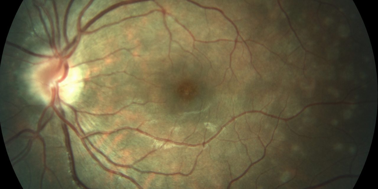
A 28-year-old female presented to the ophthalmology clinic with a sudden onset of blurred vision and photopsia in her left eye, reporting a grayish spot in her central vision.
She denied recent illness, trauma, or changes in medication. Clinical examination revealed visual acuity of 20/40 in the left eye, normal pupillary reaction, intraocular pressure within normal limits, and unremarkable anterior segment examination.
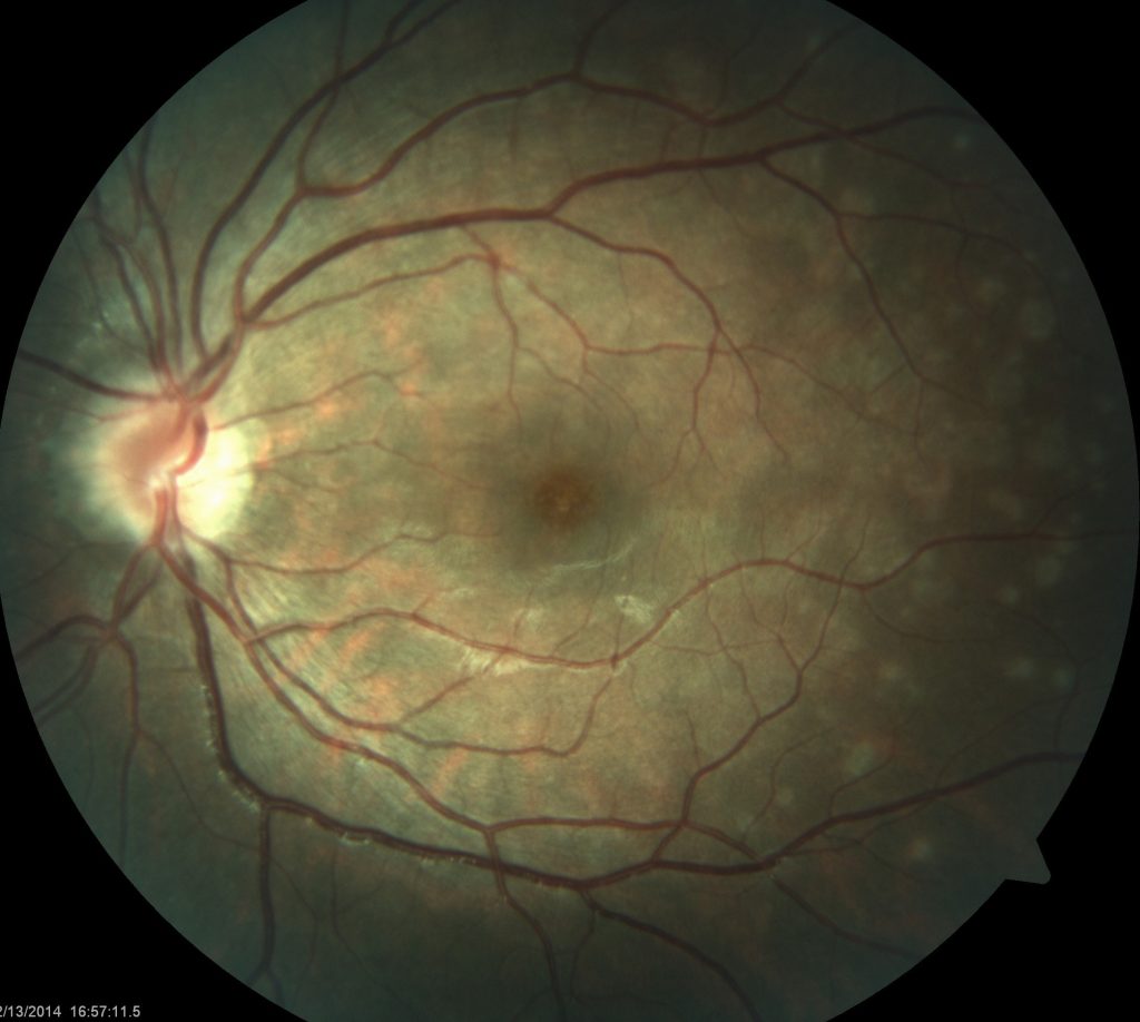
A smartphone fundus examination of the left eye showed multiple small, white dots at the level of the outer retina, primarily in the posterior pole, with mild disc edema.
Diagnostic evaluation, including fluorescein angiography, optical coherence tomography (OCT), and visual field testing, revealed hyperfluorescent spots on angiography, outer retinal disruption, hyper-reflective spots on OCT, and a mild central scotoma in the left eye.
Blood tests, including CBC, ESR, CRP, ANA, and ACE levels, were normal. The patient was diagnosed with Multiple Evanescent White Dot Syndrome (MEWDS) based on the clinical presentation and diagnostic findings.
DISEASE
Multiple Evanescent White Dot Syndrome (MEWDS) is one of the diagnoses within the family of White Dot/ White Spot Syndromes, first described by Jampol L. M and colleagues.
Within this diagnostic group are MEWDS, Acute Posterior Multifocal Placoid Pigment Epitheliopathy (APMPEE), Multifocal Choroiditis and Panuveitis (MCP), Punctate Inner Choroiditis (PIC), and Birdshot Chorioretinopathy.
The White Dot Syndromes produce yellow-white retinal lesions classically located at the retinal pigment epithelium or outer retina and are found primarily in young adults.
Symptoms of Multiple Evanescent White Dot Syndrome (MEWDS) include unilateral blurred vision, visual field loss, photopsias, and floaters. The various diagnoses are differentiated by history, appearance, laterality, and fluorescein angiogram findings.
The typical patient with MEWDS is a healthy middle-aged female aged 15-50. There is a gender disparity as women are affected with MEWDS four times more often than men.
Roughly 30% of patients have experienced an associated viral prodrome. Patients present with acute, painless, unilateral changes in vision.
They may notice photopsias, dyschromatopsia, or a temporal or paracentral scotoma. The majority of the cases have unilateral involvement, but bilateral MEWDS has been described rarely and is usually asymmetric.
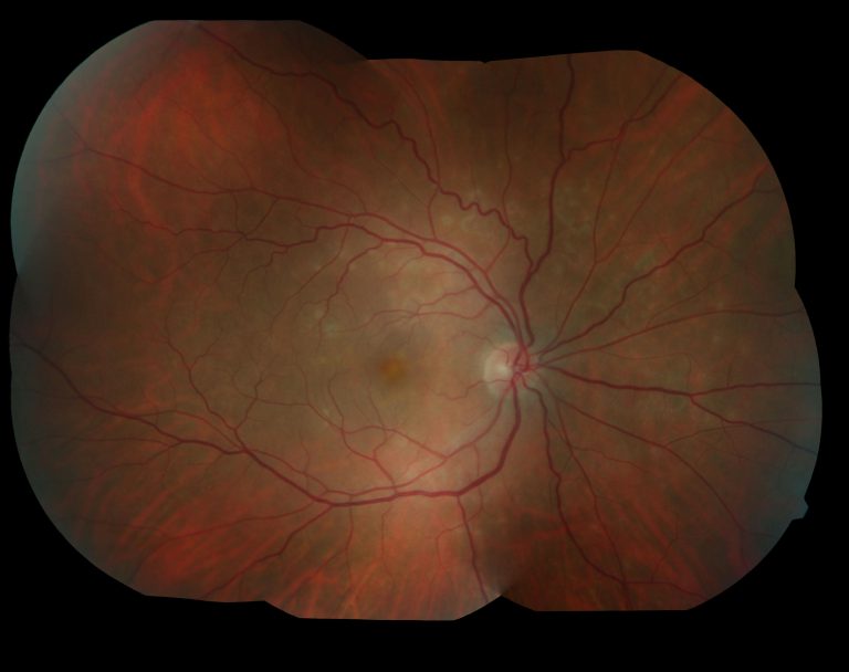
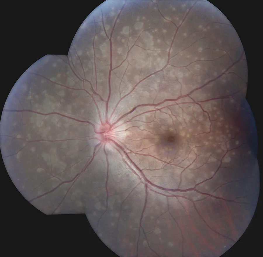
On the fundus exam, one sees flat, multifocal, grey-white lesions (100-200 microns), appearing to reach as deep as the RPE.
Typically lesions are found outside the fovea in the posterior pole. A characteristic finding in Multiple Evanescent White Dot Syndrome (MEWDS) is an orange-yellow fovea with granularity.
One might also note optic disc edema, mild vitritis (usually posterior vitreous cells), mild anterior chamber flare, a relative afferent pupillary defect, and an enlarged blind spot. Sheathing of retinal veins and superficial retinal hemorrhages are rarely seen.

Fundus Explorer Pro
Photograph the retinal findings described here with the phone already in your pocket — 22 D optics and built-in illumination in one handheld unit.
From Choroida — the team behind this siteMANAGEMENT
Frequently, a viral prodrome is elicited from the patient, though an infectious etiology is yet to be elucidated. The exact pathogenesis is unknown.
Imaging modalities such as ICGA and SD-OCT suggest the disease is caused by vaso-occlusive problems of choriocapillaris producing transient ischemia in the outer retina and reversible damage to the outer segments of the photoreceptors.
Early and late hyperfluorescent lesions on the FA may represent reactive retinal vasodilatation and exudation caused by hypo or non-perfusion of choriocapillaris.
Since Multiple Evanescent White Dot Syndrome (MEWDS) is a self-limited disease, with almost all patients regaining good visual acuity within 3-9 weeks, no treatment is recommended.
Photopsias and scotomata gradually resolve and the lesions will disappear and may be replaced by mild pigment mottling or chorioretinal scarring.
Multiple Evanescent White Dot Syndrome (MEWDS) typically is a self-limited disease, however, patients with MEWDS may have persistent blind spot enlargement.
While it is uncommon, 10% of patients may also experience a recurrence. The prognosis is relatively good for these patients.


Document what you see
Two smartphone imaging tools built for everyday clinic use — one for the slit lamp, one for the fundus.
From Choroida — the team behind this siteREFERENCES
- Jampol, Lee M. “Multiple Evanescent White Dot Syndrome: I. Clinical Findings.” Archives of Ophthalmology 102, no. 5 (May 1, 1984): 671. doi:10.1001/archopht.1984.01040030527008.
- Tavallali A, Yannuzzi LA. MEWDS, Common Cold of the Retina. J Ophthalmic Vis Res. 2017 Apr-Jun;12(2):132-134. doi: 10.4103/jovr.jovr_241_16.
- Lavigne, Luciana Castro, David Leonardo Cruvinel Isaac, José Osório Duarte Júnior, and Marcos Pereira de Avila. “Transient Spectral Domain Optical Coherence Tomography Findings in Classic MEWDS: A Case Report.” Arquivos Brasileiros De Oftalmologia 77, no. 3 (June 2014): 185–87.
- Nguyen, My Hanh T., Andre J. Witkin, Elias Reichel, Tony H. Ko, James G. Fujimoto, Joel S. Schuman, and Jay S. Duker. “Microstructural Abnormalities in MEWDS Demonstrated by Ultrahigh Resolution Optical Coherence Tomography.” Retina (Philadelphia, Pa.) 27, no. 4 (May 2007): 414–18. doi:10.1097/01.iae.0000246676.88033.25.
- Papasavvas I, Mantovani A, Tugal-Tutkun I, Herbort CP Jr. Multiple evanescent white dot syndrome (MEWDS): update on practical appraisal, diagnosis and clinicopathology; a review and an alternative comprehensive perspective. J Ophthalmic Inflamm Infect. 2021 Dec 18;11(1):45. doi: 10.1186/s12348-021-00279-7. PMID: 34921620; PMCID: PMC8684571.
