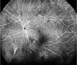
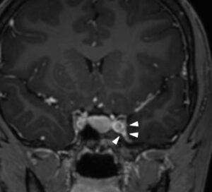
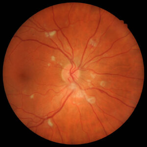
Orbital pseudotumor is a noninfectious inflammation of the orbit with no identifiable local or systemic cause.
Most specialists now call it idiopathic orbital inflammation, which typically presents with acute pain, proptosis and periorbital swelling. However, it is a diagnosis of exclusion, and several serious diseases look identical at first.
What Orbital Pseudotumor Is
Histology shows a polymorphous infiltrate of lymphocytes, plasma cells and macrophages with variable fibrosis. The trigger remains unknown, although an immune-mediated process seems likely.
Many orbital series place it among the common causes of orbital disease, after thyroid eye disease. Most patients are adults, while children form a smaller and distinct group.
Anatomical Subtypes
The inflamed tissue defines the subtype.
- Dacryoadenitis: lacrimal gland, the most frequent site
- Myositis: one or more extraocular muscles
- Anterior: globe, sclera and anterior orbit
- Apical: orbital apex, with early optic nerve risk
- Perineuritis: optic nerve sheath
- Diffuse: several compartments at once
Location predicts symptoms, and it also guides where to biopsy.
All-fit Slit-Lamp Adapter
Record and share exactly what you see at the slit lamp. One adapter fits any slit lamp or surgical microscope — and any smartphone.
From Choroida — the team behind this siteSymptoms of Orbital Pseudotumor
Pain is the leading complaint and usually begins abruptly over hours to days. Patients then notice swelling, redness and a feeling that the eye is pushed forward.
Pain on eye movement suggests myositis, and diplopia, blurred vision or tearing may follow, depending on the site.
Exam Signs at the Slit Lamp
The external exam often shows lid edema, erythema, chemosis and conjunctival injection. Lacrimal gland disease produces an S-shaped upper lid and superotemporal tenderness.
Next, measure proptosis with an exophthalmometer and test ductions for restriction. Fundus examination may reveal disc edema or choroidal folds in posterior disease.
Optic Nerve Assessment in Orbital Pseudotumor
Check acuity, color vision and pupils at every visit. A relative afferent pupillary defect signals optic nerve compromise and demands urgent treatment.
Apical and perineuritic forms carry the highest risk because the apex leaves little room.
Imaging Orbital Pseudotumor
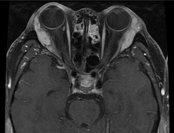
Contrast-enhanced MRI or CT of the orbits confirms the location and extent. Typical findings include an ill-defined enhancing infiltrate, a lacrimal gland molding around the globe, or a thickened muscle.
In myositis, the tendon is usually involved, while thyroid eye disease classically spares it. Bone erosion points toward malignancy or another process.
B-scan may also show fluid in Tenon’s space when the posterior sclera is inflamed (see posterior scleritis imaging).
Laboratory Workup
Basic tests include a complete blood count, inflammatory markers and thyroid function. Then add ANCA, angiotensin-converting enzyme and serum IgG4 when the history fits.
A normal panel does not exclude systemic disease. Therefore, ask about sinus symptoms, cough, weight loss and night sweats.
Mimics to Exclude in Orbital Pseudotumor
- Orbital cellulitis, especially with sinusitis and fever (see orbital cellulitis)
- Thyroid eye disease, usually with lid retraction and less pain
- Lymphoma, which may respond briefly to steroids
- Metastasis and other orbital tumors
- Granulomatosis with polyangiitis and sarcoidosis
- IgG4-related ophthalmic disease
For a broader approach, review the proptosis differential diagnosis.
When to Biopsy
Consider biopsy for atypical cases, poor steroid response, recurrence, or an accessible lacrimal lesion. Consensus criteria give histology a central role in excluding specific diseases.
Ideally, take tissue before steroids, because treatment can blur the pathology. Apical lesions are harder to reach, so surgeons weigh surgical risk against an empirical trial.
Steroid Response in Orbital Pseudotumor
Oral corticosteroids are first-line therapy, and pain often improves dramatically within 24 to 48 hours. However, a rapid response does not prove the diagnosis, since lymphoma can also improve.
Taper slowly over weeks, because rapid withdrawal often triggers relapse.
Ocular Risks of Steroid Therapy
Prolonged courses can raise intraocular pressure and promote posterior subcapsular cataract. Therefore, check pressure at follow-up visits.
Also monitor glucose, blood pressure and bone health with the primary physician.
Recurrent and Resistant Disease
Relapse is common during taper or after stopping steroids. Steroid-sparing options include methotrexate, mycophenolate mofetil and rituximab.
Low-dose orbital radiotherapy suits selected patients. Repeat biopsy at recurrence, as lymphoma or IgG4-related disease may declare itself later.
Orbital Pseudotumor in Children
Children present with bilateral disease more often than adults, and they may also show fever, headache or eosinophilia. Several reports describe more frequent uveitis and disc edema in this group.
First, rule out cellulitis, which is far more common at this age.
Counseling Patients
Explain that the condition is inflammatory, once the workup rules out infection and tumor. Next, set expectations: steroids act quickly, but the taper is long.
Tell patients to return at once if vision drops or pain returns. Finally, discuss relapse risk and the need for follow-up.
Prognosis of Orbital Pseudotumor
Most patients respond well and keep good vision. However, sclerosing forms respond poorly and may leave restricted motility or optic neuropathy.
As a result, long follow-up helps detect late systemic disease.
Pitfalls to Avoid
- Do not start steroids before excluding infection
- Never treat a rapid steroid response as diagnostic
- Avoid abrupt tapering
- Biopsy recurrent or atypical lesions
- Recheck the optic nerve at every visit


Document what you see
Two smartphone imaging tools built for everyday clinic use — one for the slit lamp, one for the fundus.
From Choroida — the team behind this siteReferences
- Yuen SJ, Rubin PA. Idiopathic orbital inflammation: distribution, clinical features, and treatment outcome. Arch Ophthalmol. 2003;121:491-499.
- Mombaerts I, Goldschmeding R, Schlingemann RO, Koornneef L. What is orbital pseudotumor? Surv Ophthalmol. 1996;41:66-78.
- Mombaerts I, Bilyk JR, Rose GE, et al. Consensus on diagnostic criteria of idiopathic orbital inflammation using a modified Delphi approach. JAMA Ophthalmol. 2017;135:769-776.
- Gordon LK. Orbital inflammatory disease: a diagnostic and therapeutic challenge. Eye (Lond). 2006;20:1196-1206.
- American Academy of Ophthalmology. Basic and Clinical Science Course, Section 7: Oculofacial Plastic and Orbital Surgery. San Francisco: AAO; 2023.

Writes and reviews the clinical articles on Educate. Every post is checked against peer-reviewed sources before it goes live.