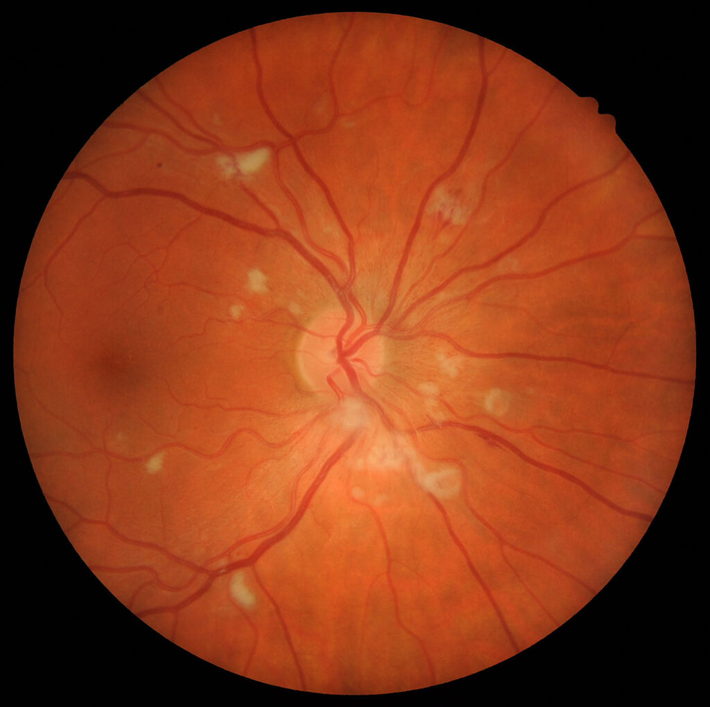
Soft exudates are small white retinal lesions, now usually called cotton wool spots.
Hard exudates, in contrast, are yellow lipid deposits that reflect chronic vascular leakage, so separating them at the first fundus examination directs the workup and follow-up.
Soft Exudates vs Hard Exudates at a Glance
- Layer: soft exudates lie in the nerve fiber layer, while hard exudates collect mainly in the outer plexiform layer
- Appearance: soft exudates look white and fluffy, whereas hard exudates look yellow, waxy and sharply defined
- Mechanism: soft exudates follow focal ischemia, while hard exudates follow chronic leakage
- Course: soft exudates usually fade within weeks, while hard exudates may persist for months
Why Soft Exudates Is a Misleading Name
The term suggests leaked material, yet these lesions contain no true exudate. Instead, occlusion of a precapillary arteriole interrupts axoplasmic flow in the nerve fiber layer, and axoplasmic debris then accumulates within swollen axons.
Fundus Explorer Pro
Photograph the retinal findings described here with the phone already in your pocket — 22 D optics and built-in illumination in one handheld unit.
From Choroida — the team behind this siteHow Soft Exudates Look on Examination
The examiner sees a small, white, superficial patch with feathery edges, and because the lesion sits in the inner retina, it can obscure the underlying vessels. Patients are often asymptomatic, although a spot near fixation may produce a small scotoma.
How Hard Exudates Form
Hard exudates begin when damaged capillaries or microaneurysms leak plasma into the retina. The water component is then resorbed faster than the lipid and protein, so lipoprotein and lipid-laden macrophages remain within the outer retina. Deposits may form dots, plaques, or a circinate ring around a leaking point (see hard exudates).
Soft Exudates on OCT
Spectral-domain OCT shows focal hyperreflective thickening of the inner retina. Later, the affected nerve fiber layer often thins as the spot resolves.
Hard Exudates on OCT
Hard exudates appear as dense hyperreflective foci, mainly in the outer retinal layers, and they frequently cast a posterior shadow. They often accompany intraretinal cysts, and therefore OCT also reveals the macular edema that maintains the deposits.
Systemic Causes of Soft Exudates
Diabetes and hypertension account for many cases seen in routine practice, but a spot without either condition needs a wider search. In their classic series, Brown and colleagues linked isolated spots to serious underlying disease. Consider these associations, then tailor testing to age and symptoms:
- Retinal vein occlusion and carotid or cardiac emboli
- Lupus, giant cell arteritis and other vasculitic disease
- HIV retinopathy, anemia and leukemia
Ocular Causes of Hard Exudates
Diabetic macular edema is the most common setting in clinical practice, and retinal vein occlusion and retinal arterial macroaneurysm can also produce circinate lipid rings. Finally, younger patients with massive peripheral lipid need evaluation for Coats disease.
What Soft Exudates Mean Clinically
These lesions indicate recent focal ischemia of the inner retina, and in diabetic eyes, multiple spots may accompany other signs of worsening nonperfusion. In hypertensive patients, they support a more advanced retinopathy grade and warrant prompt blood pressure assessment.
What Hard Exudates Mean Clinically
Hard exudates mark chronic leakage, and their threat depends largely on location, with deposits near the foveal center raising concern for clinically significant macular edema. The ETDRS also associated elevated serum lipids with greater retinal hard exudate, and subfoveal lipid can progress to fibrosis, which limits visual recovery.
Managing Soft Exudates
No direct retinal treatment is needed for the spot itself, so management instead targets the underlying vascular, inflammatory or hematologic disease. Tell patients that the spot is a warning sign, while any scotoma may persist.
Managing Hard Exudates
Treatment aims to stop leakage at its source, usually with anti-VEGF therapy for diabetic macular edema, and focal laser or corticosteroids may also suit selected eyes. However, deposits clear slowly, and established subfoveal scarring will not reverse. Therefore, counsel patients that a clearer fundus may not mean better vision.
Conditions That Mimic Soft Exudates
Myelinated nerve fibers also look white, but they remain stable and show feathered margins, whereas retinitis produces denser, progressive whitening, often with vitritis. Soft drusen sit beneath the retina (see soft versus hard drusen), and for a full disease review, read our article on cotton wool spots.
Follow-Up for Soft Exudates and Hard Exudates
Rephotograph soft exudates after several weeks and confirm that older lesions are fading, and new spots should prompt a repeat check of blood pressure and glucose. For hard exudates, pair color photographs with OCT and record their distance from the fovea.
Pitfalls to Avoid
- Do not assume every white lesion is lipid, because color and depth matter
- Never attribute an isolated spot to age without a systemic evaluation
- Do not treat lipid alone while ignoring active macular edema


Document what you see
Two smartphone imaging tools built for everyday clinic use — one for the slit lamp, one for the fundus.
From Choroida — the team behind this siteReferences
- Brown GC, Brown MM, Hiller T, et al. Cotton-wool spots. Retina. 1985;5:206-214.
- McLeod D. Why cotton wool spots should not be regarded as retinal nerve fibre layer infarcts. Br J Ophthalmol. 2005;89:229-237.
- Schmidt D. The mystery of cotton-wool spots: a review of recent and historical descriptions. Eur J Med Res. 2008;13:231-266.
- Chew EY, Klein ML, Ferris FL, et al. Association of elevated serum lipid levels with retinal hard exudate in diabetic retinopathy: ETDRS report 22. Arch Ophthalmol. 1996;114:1079-1084.
- Wong TY, Mitchell P. Hypertensive retinopathy. N Engl J Med. 2004;351:2310-2317.

Writes and reviews the clinical articles on Educate. Every post is checked against peer-reviewed sources before it goes live.