Retinal folds are creases in the neurosensory retina, while the underlying retinal pigment epithelium (RPE) usually stays flat.
They arise from abnormal development, surface traction or shifts during retinal reattachment.
Therefore, the patient’s age and surgical history narrow the cause quickly.
OCT then confirms which layer is folded.
What Retinal Folds Are
The retina buckles when a force drags it sideways or when fluid shifts its position.
Folds may involve the full thickness, the inner layers or the outer layers alone.
For example, an epiretinal membrane wrinkles the inner retina.
Symptoms of Retinal Folds
Many peripheral folds cause no symptoms.
However, a fold crossing the fovea commonly produces metamorphopsia and reduced acuity.
Children with congenital folds may present with strabismus or nystagmus.
Adults may notice distortion or double vision after detachment repair.
Fundus Explorer Pro
Photograph the retinal findings described here with the phone already in your pocket — 22 D optics and built-in illumination in one handheld unit.
From Choroida — the team behind this siteCongenital Retinal Folds
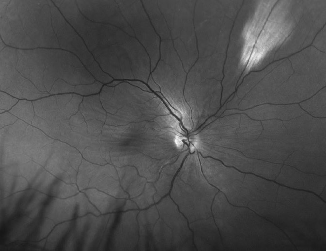
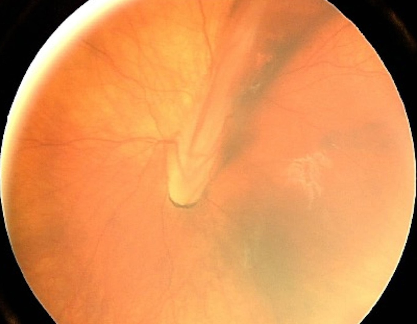
These folds typically run from the optic disc toward the temporal periphery, often carrying retinal vessels.
Older texts call this appearance a falciform fold.
The peripheral end may attach to retrolental tissue.
Vision depends on macular involvement.
Conditions Behind Congenital Folds
Familial exudative vitreoretinopathy (FEVR) and retinopathy of prematurity can create folds through peripheral fibrovascular traction.
Persistent fetal vasculature and Norrie disease can also produce them.
Therefore, examine both peripheral retinas and ask about prematurity and family history.
Because FEVR may be subtle in relatives, widefield angiography of family members can help.
A Benign Peripheral Mimic
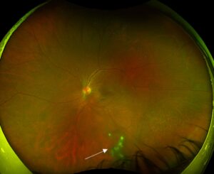
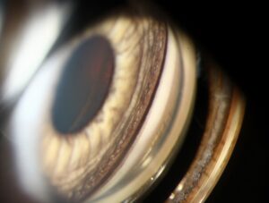
Meridional folds are small radial elevations near the ora serrata (see meridional folds).
They usually align with a dentate process and rarely cause problems.
However, check the posterior tip for a small retinal break.
Tractional Retinal Folds
Epiretinal membranes contract tangentially and wrinkle the inner retina around the macula.
OCT shows a hyperreflective surface membrane and an undulating inner retinal contour.
Meanwhile, the RPE line stays smooth.
Proliferative Membranes and Star Folds
Proliferative vitreoretinopathy creates stiff star folds, usually after detachment or repair.
Diabetic fibrovascular membranes can also crease the retina along the arcades (see tractional retinal detachment).
Therefore, look for subretinal fluid, breaks and progression between visits.
Postoperative Retinal Folds
Full-thickness folds can follow scleral buckling, pneumatic retinopexy or vitrectomy with gas.
Reported associations include bullous detachment and residual subretinal fluid.
A fold through the fovea needs early surgical assessment, because fixed folds may limit recovery.
Outer Retinal Folds After Repair
OCT may show undulating outer retinal layers after macula-off repair.
These folds often flatten over weeks to months.
Therefore, observation usually suits small folds that spare the fovea.
Serial OCT then documents recovery of the external limiting membrane and ellipsoid zone.
Retinal Displacement After Surgery
The retina may reattach slightly away from its original position, even without a visible fold.
Shiragami and colleagues described this unintentional displacement after vitrectomy for rhegmatogenous detachment.
Autofluorescence can reveal parallel hyperautofluorescent lines beside the retinal vessels.
Patients may report distortion, so a normal-looking fundus does not end the evaluation.
Retinal Folds Versus Choroidal Folds on OCT
Start with the RPE and Bruch membrane on the OCT scan.
In retinal folds, this band stays flat while the neurosensory retina creases above it.
In choroidal folds, the RPE and Bruch membrane undulate together (see choroidal folds).
Finally, scan perpendicular to the lines, since a parallel scan can hide the undulation.
Fundus and Angiography Clues
Choroidal folds usually appear as broad parallel dark and light bands across the posterior pole.
Retinal folds tend to look finer and often radiate from a point of traction.
Angiography also shows alternating hyperfluorescent and hypofluorescent bands in choroidal folds.
When the Choroid Is Involved
Hyperopia, hypotony, posterior scleritis, orbital masses and thyroid eye disease are recognized causes.
Unilateral folds call for orbital and scleral evaluation.
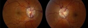
Also, inspect the disc, because papilledema can produce peripapillary wrinkles.
Managing Retinal Folds
Treatment follows the mechanism.
Congenital folds rarely unfold, so care focuses on refraction, amblyopia therapy and active peripheral disease.
Vitrectomy with membrane peeling can relieve tractional distortion; however, some metamorphopsia may persist, and surgery carries cataract and retinal break risks.
Early reoperation may unfold a postoperative macular fold, although the evidence comes mainly from case series.
Counseling the Patient
Explain which layer is folded and whether the fold threatens vision.
Surgery can flatten the anatomy without restoring normal sight.
Also, ask patients to report new distortion, floaters or field loss.
Pitfalls to Avoid
- Do not label every line on OCT as a retinal fold before checking the RPE
- Avoid delaying assessment of a new postoperative fold through the fovea
- Never skip the peripheral exam in a child with a fold


Document what you see
Two smartphone imaging tools built for everyday clinic use — one for the slit lamp, one for the fundus.
From Choroida — the team behind this siteReferences
- Ranchod TM, Ho LY, Drenser KA, Capone A Jr, Trese MT. Clinical presentation of familial exudative vitreoretinopathy. Ophthalmology. 2011;118:2070-2075.
- Heimann H, Bopp S. Retinal folds following retinal detachment surgery. Ophthalmologica. 2011;226(Suppl 1):18-26.
- Shiragami C, Shiraga F, Yamaji H, et al. Unintentional displacement of the retina after standard vitrectomy for rhegmatogenous retinal detachment. Ophthalmology. 2010;117:86-92.
- Olsen TW, Palejwala NV, Lee LB, Bergstrom CS, Yeh S. Chorioretinal folds: associated disorders and a related maculopathy. Am J Ophthalmol. 2014;157:1038-1047.
- Leahey AB, Brucker AJ, Wyszynski RE, Shaman P. Chorioretinal folds: a comparison of unilateral and bilateral cases. Arch Ophthalmol. 1993;111:357-359.

Writes and reviews the clinical articles on Educate. Every post is checked against peer-reviewed sources before it goes live.