OCT angiography artifacts are errors in flow images that can create false vessels, hide real ones, or distort capillary measurements.
The technology detects motion contrast, so anything that moves or changes signal may appear as flow. Clinicians who read these images need to know the common artifacts and their causes.
Why OCT Angiography Artifacts Occur
OCT angiography compares repeated scans of the same location, and it assigns flow to areas where the signal changes, while eye movement, tissue changes, and software processing also change the signal.
Spaide and colleagues reviewed these errors, and they are present in a large proportion of scans. Therefore, a careful reading always includes the structural OCT (see OCT angiography).
Projection OCT Angiography Artifacts
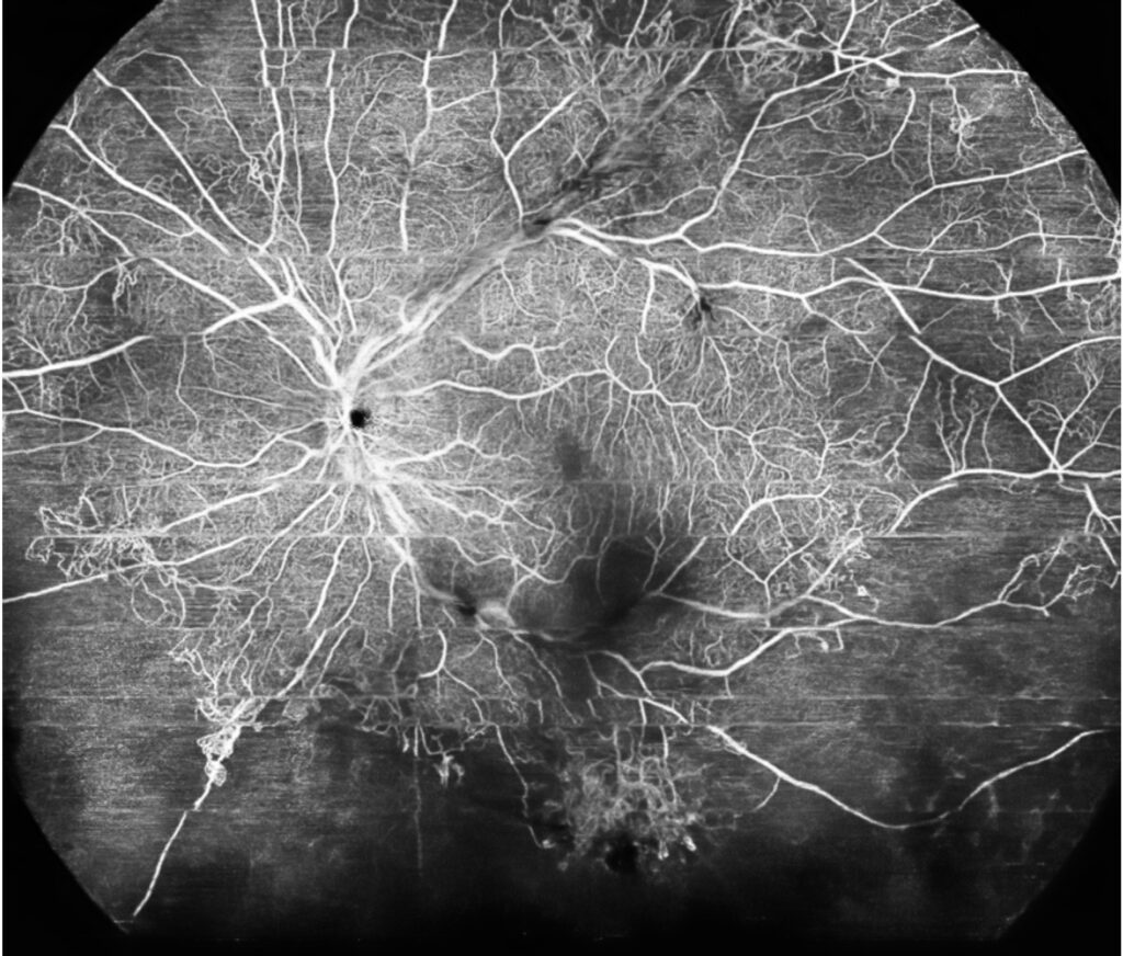
Flow in superficial vessels casts a shadow in deeper layers, and the software may show copies of those vessels in the outer retina, which can mimic choroidal neovascularization in the outer retina slab.
Projection removal algorithms reduce the problem but may leave residual artifacts or erase real vessels, so check the B-scan with flow overlay to confirm that real flow lies in the layer (see macular neovascularization types).
Fundus Explorer Pro
Photograph the retinal findings described here with the phone already in your pocket — 22 D optics and built-in illumination in one handheld unit.
From Choroida — the team behind this siteMotion and Blink Artifacts
Eye movement creates white lines, doubled vessels, and discontinuities, and tracking and motion correction software reduce them, but unsteady patients still produce poor scans. Repeat the scan, or choose a smaller field, when motion is a problem.
Segmentation Artifacts
Errors in layer boundaries shift the slab, and the image then shows the wrong tissue. In diabetic and myopic eyes, thinning and distortion increase these errors, and manual correction of boundaries is time consuming, but it prevents false conclusions (see retinal capillary nonperfusion).
Shadow and Signal OCT Angiography Artifacts
- Vitreous floaters and opacities that block signal and produce dark areas
- Hemorrhage or exudate that shadows deeper layers
- Low signal strength that makes the capillaries look sparse
- Media opacities that reduce the entire image quality
Dark areas that match a shadow on the structural image are not nonperfusion.
Avoiding OCT Angiography Artifacts
Use a good signal strength, and check that the image quality meets the manufacturer’s threshold. Compare slabs with the B-scan, and avoid reading vessel density from a single poor scan. Use the same device and protocol for follow-up, since values differ between instruments.
An Example Case
A 66-year-old man has a suspected neovascular membrane after cataract surgery, and the outer retina slab shows a tangle of vessels, with similar patterns in the superficial plexus above it.
On the B-scan with flow overlay, the signal in the outer retina turns out to be a projection of the superficial vessels, with no true flow beneath the RPE, so the clinician avoids unnecessary treatment and schedules a repeat scan.
Questions Clinicians Ask
Clinicians ask whether projection removal solves the problem, and the answer is that it helps but may erase real signal. They also ask how often artifacts occur, and large studies report them in a substantial portion of scans. Because artifacts vary by device, learn the behavior of your instrument.
Pitfalls to Avoid
- Diagnosing a membrane from the en face slab alone
- Reading dark patches as nonperfusion without checking the structural image
- Comparing vessel density between different devices
- Using a poor-quality scan for quantitative analysis
Looking Ahead
New algorithms will improve projection removal and motion correction, and wider fields and faster scanning will reduce artifacts from eye movement. Therefore, the next generation of devices may simplify interpretation.
Practical Points
Confirm the finding on at least two views, such as the en face slab and the B-scan with flow. Then repeat the scan when the finding changes the treatment plan, and document the image quality score in the record.
Quantitative OCT Angiography and Artifacts
Vessel density, foveal avascular zone area, and perfusion indices all depend on image quality. Therefore, exclude scans with low signal or visible motion before analysis. Use the same device and software version for follow-up, and report the signal strength in studies, since it affects the results.
Teaching Points
Always read the structural B-scan first, then the flow overlay, and then the en face slab, because this habit prevents most misreadings. Because artifacts are common, teach residents to question every unusual vascular pattern.
Remind the team that image quality changes with pupil size, so dilate when the media allow, and because scans take longer than structural OCT, schedule enough time.


Document what you see
Two smartphone imaging tools built for everyday clinic use — one for the slit lamp, one for the fundus.
From Choroida — the team behind this siteReferences
- Spaide RF, Fujimoto JG, Waheed NK. Image artifacts in optical coherence angiography. Retina. 2015;35:2163-2180.
- Ghasemi Falavarjani K, Al-Sheikh M, Akil H, Sadda SR. Image artefacts in swept-source optical coherence tomography angiography. Br J Ophthalmol. 2017;101:564-568.
- de Carlo TE, Romano A, Waheed NK, Duker JS. A review of optical coherence tomography angiography (OCTA). Int J Retina Vitreous. 2015;1:5.
- American Academy of Ophthalmology. Basic and Clinical Science Course, Section 12: Retina and Vitreous. AAO; current edition.

Writes and reviews the clinical articles on Educate. Every post is checked against peer-reviewed sources before it goes live.