Epithelial downgrowth is the invasion of surface epithelium into the anterior chamber after intraocular surgery or penetrating trauma.
The cells line the posterior cornea, iris and angle as a sheet, or they form a cyst. It is uncommon today, yet late diagnosis can still cost the eye, so anyone who follows postoperative eyes should recognize its signs.
What Epithelial Downgrowth Is
Conjunctival or corneal epithelium normally stops at the wound surface. However, a gaping or poorly apposed wound gives the cells a path inward, and once inside, they spread as a thin membrane over the endothelium, trabecular meshwork and iris.
Sheet, Cystic and Fibrous Forms
The sheetlike form is the classic and most aggressive pattern, whereas the cystic form appears as a translucent cyst on the iris or near the wound. Fibrous ingrowth is a separate entity made of fibroblastic tissue that usually looks thicker and whiter, and histology separates the two when doubt remains.
All-fit Slit-Lamp Adapter
Record and share exactly what you see at the slit lamp. One adapter fits any slit lamp or surgical microscope — and any smartphone.
From Choroida — the team behind this siteRisk Factors for Epithelial Downgrowth
- Wound leak or delayed wound closure
- Complicated cataract surgery with vitreous incarceration
- Descemet membrane tears at the incision
- Penetrating keratoplasty and repeat grafts
- Penetrating trauma with tissue caught in the wound
Why Small Incisions Changed Epithelial Downgrowth
Large intracapsular and extracapsular incisions carried a higher risk, and clear corneal phacoemulsification has since made the condition rare. However, case reports still describe downgrowth after small incision surgery, so a small wound does not exclude the diagnosis.
Symptoms Patients Report
Many patients notice slow, painless blurring months or years after surgery, while others report redness, photophobia or ache when pressure rises. Because symptoms are often mild, the slit lamp frequently finds the problem first.
The Retrocorneal Membrane in Epithelial Downgrowth
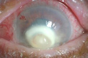
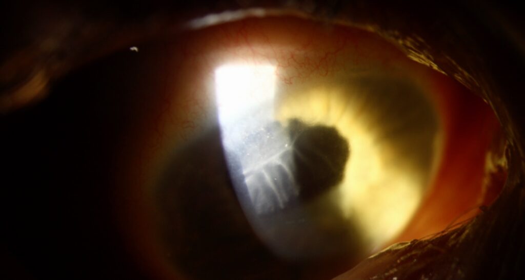
The hallmark sign is a gray, translucent membrane on the back of the cornea, and its advancing edge is sharp and often scalloped. The membrane usually starts at an old wound and then spreads centrally, and the overlying cornea may become edematous.
Other Slit-Lamp Signs
- Positive Seidel test or a filtering bleb at the wound
- Distorted or drawn-up pupil
- Loss of iris crypts under a glassy film
- Translucent cyst arising from the iris or wound
Gonioscopy and Glaucoma in Epithelial Downgrowth
Gonioscopy shows the membrane crossing the angle structures, and outflow then falls as epithelium covers the trabecular meshwork. Because of this, the glaucoma is often severe and resists drops, so unexplained pressure rise after a complicated wound deserves a careful angle exam (compare angle recession glaucoma).
The Argon Laser Test
Low-energy argon burns help map iris involvement, because normal iris stroma turns brown and contracts after a burn. However, iris covered by epithelium blanches to a fluffy white, so the surgeon can outline the affected zone before surgery.
Imaging in Epithelial Downgrowth
Anterior segment OCT can show a hyperreflective layer behind the cornea or over the iris. Specular microscopy may reveal a sharp line between normal endothelium and abnormal cells, and ultrasound biomicroscopy also defines cysts and angle involvement.
Differential Diagnosis
- Fibrous ingrowth
- Descemet membrane detachment (see Descemet membrane detachment)
- Endothelial rejection line after keratoplasty
- Iridocorneal endothelial syndrome
Epithelial ingrowth under a LASIK flap is also different, because it stays in the interface (see epithelial ingrowth after LASIK).
Surgical Treatment of Epithelial Downgrowth
Classic surgery aims to remove or destroy all invading epithelium. Surgeons excise involved iris and vitreous, then freeze the corneal membrane with cryotherapy, and they also close any fistula and repair the wound. However, this extensive procedure can damage the endothelium, and a corneal graft may follow.
Intracameral 5-Fluorouracil
Case reports describe intracameral 5-fluorouracil as a less invasive option, and the drug targets dividing epithelial cells inside the chamber. However, the evidence comes from single cases and small series, and endothelial toxicity and recurrence also remain open questions.
Controlling Pressure
Glaucoma often persists after the membrane stops advancing. Therefore, many eyes need a glaucoma drainage device, and cyclophotocoagulation is an option when visual potential is poor.
Counseling the Patient
First, explain that surface cells have grown inside the eye, and next, state that treatment aims to stop spread and preserve useful vision. Finally, set honest expectations, because the prognosis is guarded.
Follow-Up After Epithelial Downgrowth Treatment
Measure pressure and photograph the membrane edge at each visit, and also recheck the wound for leaks and the endothelium for decompensation (see endothelial cell loss).
Pitfalls to Avoid
- Do not call a gray retrocorneal line simple edema without gonioscopy
- Never ignore a persistent wound leak after surgery
- Avoid blind cyst drainage, because spilled cells may seed the chamber
- Do not assume a small incision excludes the diagnosis


Document what you see
Two smartphone imaging tools built for everyday clinic use — one for the slit lamp, one for the fundus.
From Choroida — the team behind this siteReferences
- Weiner MJ, Trentacoste J, Pon DM, Albert DM. Epithelial downgrowth: a 30-year clinicopathological review. Br J Ophthalmol. 1989;73:6-11.
- Maumenee AE, Paton D, Morse PH, Butner R. Review of 40 histologically proven cases of epithelial downgrowth following cataract extraction and suggested surgical management. Am J Ophthalmol. 1970;69:598-603.
- Vargas LG, Vroman DT, Solomon KD, et al. Epithelial downgrowth after clear cornea phacoemulsification: report of two cases and review of the literature. Ophthalmology. 2002;109:2331-2335.
- Lai MM, Haller JA. Resolution of epithelial ingrowth in a patient treated with 5-fluorouracil. Am J Ophthalmol. 2002;133:562-564.
- American Academy of Ophthalmology. Basic and Clinical Science Course, Section 10: Glaucoma. San Francisco: AAO; 2023.
Test yourself
A few questions straight from this article.
-
What is epithelial downgrowth?
Epithelial downgrowth is the invasion of conjunctival or corneal surface epithelium into the anterior chamber through a poorly apposed wound after intraocular surgery or penetrating trauma. -
Which form of epithelial downgrowth is the classic and most aggressive pattern?
The sheetlike form is the classic and most aggressive pattern, while the cystic form appears as a translucent cyst on the iris or near the wound. -
How does fibrous ingrowth differ from epithelial downgrowth?
Fibrous ingrowth is a separate entity of fibroblastic tissue that usually looks thicker and whiter; histology separates the two when doubt remains. -
Which surgical event is a risk factor for epithelial downgrowth?
Risk factors include wound leak or delayed closure, complicated cataract surgery with vitreous incarceration, Descemet tears, penetrating keratoplasty and penetrating trauma. -
Why has epithelial downgrowth become rare?
Large intracapsular and extracapsular incisions carried more risk; clear corneal phaco has made it rare, though downgrowth still occurs after small incisions. -
What is the hallmark slit-lamp sign of epithelial downgrowth?
The hallmark is a grey, translucent membrane on the back of the cornea with a sharp, often scalloped advancing edge that starts at an old wound and spreads centrally. -
Why is the glaucoma of epithelial downgrowth often severe and resistant to drops?
Gonioscopy shows the membrane crossing the angle, and outflow falls as epithelium covers the trabecular meshwork, so the glaucoma is often severe and drop-resistant. -
In the argon laser test for epithelial downgrowth, how does epithelium-covered iris respond?
Normal iris stroma turns brown and contracts after a low-energy burn, whereas iris covered by epithelium blanches fluffy white, mapping the involved zone before surgery. -
How is the corneal membrane treated in classic surgery for epithelial downgrowth?
Classic surgery excises involved iris and vitreous, freezes the corneal membrane with cryotherapy, closes any fistula and repairs the wound; a corneal graft may follow. -
Which pressure-lowering surgery do many eyes with epithelial downgrowth need?
Glaucoma often persists after the membrane stops advancing, so many eyes need a glaucoma drainage device, with cyclophotocoagulation an option when visual potential is poor.

Writes and reviews the clinical articles on Educate. Every post is checked against peer-reviewed sources before it goes live.