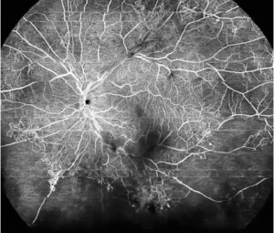
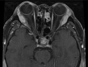
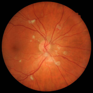
Tolosa-Hunt syndrome is painful ophthalmoplegia caused by idiopathic granulomatous inflammation of the cavernous sinus or superior orbital fissure.
It is rare, yet it appears in almost every differential of a painful third, fourth or sixth nerve palsy, but it is a diagnosis of exclusion.
What Causes Tolosa-Hunt Syndrome
Tolosa described granulomatous tissue around the carotid siphon at autopsy in 1954, and then Hunt and colleagues characterized the clinical syndrome and its steroid response in 1961. The trigger remains unknown, so most authors regard it as a nonspecific, presumed autoimmune inflammation.
Typical Presentation
Patients usually report a severe, boring pain behind or around one eye, which often precedes diplopia by several days. Then ptosis or double vision follows on the same side, and onset usually falls in adult life, with both sexes developing it.
All-fit Slit-Lamp Adapter
Record and share exactly what you see at the slit lamp. One adapter fits any slit lamp or surgical microscope — and any smartphone.
From Choroida — the team behind this siteExamination Signs in Tolosa-Hunt Syndrome
The cavernous sinus packs several structures into a small space, so combined deficits are common.
- Ipsilateral third, fourth or sixth nerve palsy, alone or combined
- Reduced forehead sensation from first trigeminal division involvement
- Oculosympathetic paresis, with a small pupil and mild ptosis
- Occasional optic neuropathy when inflammation reaches the orbital apex
Localizing the Lesion
A third nerve palsy combined with a sixth nerve palsy points to the cavernous sinus, and V1 sensory loss supports that site, while optic nerve dysfunction moves the lesion forward to the orbital apex. In contrast, an isolated adduction deficit with abducting nystagmus suggests a brainstem lesion (see internuclear ophthalmoplegia).
Diagnostic Criteria for Tolosa-Hunt Syndrome
The ICHD-3 criteria require unilateral headache and ipsilateral ocular motor nerve paresis, along with granulomatous inflammation shown on MRI or biopsy. The headache must precede the paresis by up to two weeks or accompany it. The pain must lie around the ipsilateral brow and eye, and no other diagnosis may explain the picture better.
Why Criteria Are Not Enough
Many tumors and infections satisfy the first parts of the definition, and lymphoma, for example, can produce identical pain, palsies and enhancement. Therefore, the last criterion carries most of the weight, so clinicians should treat the label as provisional until follow-up confirms it.
Mimics of Tolosa-Hunt Syndrome to Exclude
Exclude these before starting steroids whenever possible:
- Intracavernous carotid aneurysm
- Carotid-cavernous fistula, especially with chemosis or arterialized vessels
- Pituitary apoplexy, meningioma, lymphoma and metastasis
- Invasive fungal sinusitis such as mucormycosis in diabetic or immunosuppressed patients
- Sarcoidosis, IgG4-related disease and granulomatosis with polyangiitis
- Tuberculosis and herpes zoster ophthalmicus
- Diabetic microvascular palsy with periocular pain
Slit Lamp Clues to a Mimic
Dilated corkscrew episcleral vessels and raised intraocular pressure suggest a fistula, and similarly, proptosis and chemosis point toward orbital or vascular disease. Vesicles in the V1 dermatome suggest zoster, while a black nasal or palatal eschar suggests mucormycosis.
MRI Findings in Tolosa-Hunt Syndrome
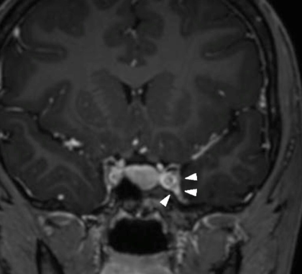
Contrast-enhanced MRI with thin cuts through the cavernous sinus and orbit is the key test, and the typical finding is enhancing soft tissue expanding the affected sinus. This tissue is usually isointense to gray matter on T1 and iso- to hypointense on T2.
It may extend into the superior orbital fissure or orbital apex, but none of these features is specific, and lymphoma or meningioma can look identical.
Laboratory Workup
Tests aim at the mimics, so a typical panel includes complete blood count, ESR, CRP, glucose and HbA1c. Many clinicians add ACE, ANCA, IgG4, syphilis serology and tuberculosis testing, and lumbar puncture helps when lymphoma, infection or carcinomatous meningitis remains possible.
Steroid Response in Tolosa-Hunt Syndrome
Systemic corticosteroids usually produce rapid pain relief, and pain often improves within about 72 hours. Cranial nerve function recovers more slowly, often over weeks, and no trial has defined the best dose or duration, so regimens vary widely. Clinicians usually taper over weeks to months under clinical and MRI follow-up.
Pitfalls of the Steroid Test
A good response does not prove the diagnosis, because lymphoma, sarcoidosis and some metastases may shrink briefly on steroids. Steroids can also worsen fungal infection or tuberculosis, so start treatment only after imaging and screening for infection. Persistent palsy beyond the expected window should prompt reassessment and biopsy.
Follow-Up and Relapse
Relapse occurs in a substantial proportion of patients, sometimes on the opposite side, and repeat MRI should show regression of the lesion. Steroid-sparing agents are an option for frequent relapses, and clinicians should also monitor intraocular pressure during long steroid courses (see steroid-induced glaucoma).
Counseling Patients With Tolosa-Hunt Syndrome
Explain that the condition is inflammatory and usually responds well to treatment, but tell patients that diplopia may take weeks to resolve. Prism or temporary occlusion can help in the meantime, and finally, ask them to return promptly if pain recurs or vision changes.
Pitfalls to Avoid
- Do not start steroids before contrast MRI
- Never accept pain relief as proof of benign disease
- Do not skip glucose testing in painful ophthalmoplegia
- Review patients with persistent deficits for biopsy


Document what you see
Two smartphone imaging tools built for everyday clinic use — one for the slit lamp, one for the fundus.
From Choroida — the team behind this siteReferences
- Headache Classification Committee of the International Headache Society. The International Classification of Headache Disorders, 3rd edition. Cephalalgia. 2018;38:1-211.
- Kline LB, Hoyt WF. The Tolosa-Hunt syndrome. J Neurol Neurosurg Psychiatry. 2001;71:577-582.
- Hunt WE, Meagher JN, LeFever HE, Zeman W. Painful ophthalmoplegia: its relation to indolent inflammation of the cavernous sinus. Neurology. 1961;11:56-62.
- Tolosa E. Periarteritic lesions of the carotid siphon with the clinical features of a carotid infraclinoidal aneurysm. J Neurol Neurosurg Psychiatry. 1954;17:300-302.
- American Academy of Ophthalmology. Basic and Clinical Science Course, Section 5: Neuro-Ophthalmology. San Francisco: AAO; 2023.

Writes and reviews the clinical articles on Educate. Every post is checked against peer-reviewed sources before it goes live.