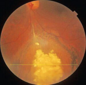
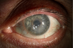
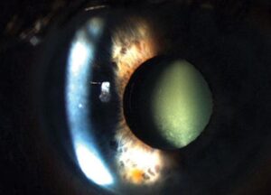
Corneal ring abscess is a circumferential stromal infiltrate with dense suppuration, and it suggests severe keratitis until proven otherwise.
It usually forms concentric to the limbus, and necrosis may follow quickly.
Therefore, manage it as a same-day emergency while you define the cause.
What Defines a Corneal Ring Abscess
The examiner sees a yellow-white annular stromal band, often with overlying epithelial loss.
A clearer zone may separate the ring from the limbus.
Patients report rapid pain, photophobia, discharge and falling vision.
Why the Ring Forms
Neutrophils enter the stroma from limbal vessels and gather along chemotactic gradients.
Meanwhile, microbial toxins and host enzymes digest collagen.
Because the host response shapes the pattern, the ring cannot identify the organism alone.
All-fit Slit-Lamp Adapter
Record and share exactly what you see at the slit lamp. One adapter fits any slit lamp or surgical microscope — and any smartphone.
From Choroida — the team behind this siteBacterial Causes of Corneal Ring Abscess
Pseudomonas aeruginosa is a classic cause, especially in contact lens wearers.
Its proteases can cause rapid stromal melting.
Bacillus species are another concern after trauma with soil or metallic material.
For example, a fast ring after penetrating injury should raise suspicion of possible endophthalmitis.
Fungal and Acanthamoeba Rings
Filamentous fungal keratitis may show an immune ring, feathery margins and satellite lesions.
By contrast, Acanthamoeba typically produces a ring infiltrate with severe pain and radial perineuritis.
Herpetic disease can also produce an immune ring.
Noninfectious Mimics of Corneal Ring Abscess
A Wessely ring is an immune deposit, and it typically lacks the necrosis of a true abscess.
Topical anesthetic abuse can also cause a ring infiltrate with negative cultures.
However, exclude infection first whenever suppuration, hypopyon or rapid progression is present.
History That Redirects the Workup
Ask about contact lens wear, water exposure, trauma, vegetable matter and recent surgery.
Also, ask directly about anesthetic drops, because patients may not volunteer this history.
Prior antibiotics matter as well, since they can lower culture yield.
Severity Signs in Corneal Ring Abscess
Record ring diameter, infiltrate depth, stromal thinning, epithelial defect size and hypopyon height.
Then, check for a positive Seidel test or a shallow anterior chamber.
Scleral extension is also a poor sign.
Triage for Corneal Ring Abscess
Same-day cornea assessment is appropriate for most patients with this finding.
Admission may help when hourly drops, poor adherence or impending perforation make outpatient care unsafe.
However, do not delay scraping when a capable clinician can obtain it immediately.
Sampling and Culture in Corneal Ring Abscess
Ideally, scrape the leading edge and ulcer base before starting antimicrobials, but never let logistics delay treatment.
First, request Gram and Giemsa stains, plus KOH or calcofluor white when fungi are possible.
Next, inoculate blood, chocolate and Sabouraud media.
Add non-nutrient agar with an E. coli overlay when Acanthamoeba is suspected.
In addition, send the lens case in lens wearers.
When Cultures Are Negative
Negative cultures do not exclude infection, particularly after antibiotic exposure.
Therefore, repeat sampling if the infiltrate advances.
Finally, a corneal biopsy can help with deep disease or repeated negative smears.
Looking Behind the Cornea
Dense opacity can hide vitritis or endophthalmitis.
Therefore, obtain a B-scan when the posterior view is poor, especially after trauma or surgery.
Treating Corneal Ring Abscess Empirically
Broad-spectrum topical therapy usually starts hourly, often using fortified agents for severe stromal disease.
A typical regimen pairs vancomycin or a cephalosporin with an aminoglycoside.
However, drops can cause surface toxicity, and they cannot restore collagen already lost.
For filamentous fungi, MUTT favored natamycin over voriconazole, particularly in Fusarium ulcers.
Steroids in Corneal Ring Abscess
The SCUT trial showed no overall vision benefit from adjunctive prednisolone in culture-proven bacterial ulcers.
Therefore, withhold steroids until the organism is known and the eye is improving.
Avoid them when fungal infection remains possible.
Perforation and Surgical Options
Progressive thinning calls for frequent review and early planning with a cornea surgeon.
Small perforations may respond to tissue adhesive and a bandage lens.
Larger defects or uncontrolled infection may need tectonic or therapeutic keratoplasty.
Corneal Ring Abscess Follow-Up
Review severe cases daily at first, then extend intervals only after clear improvement.
Reassuring signs include less pain, a smaller epithelial defect and a falling hypopyon.
Scarring can persist after cure, as discussed in this review of stromal corneal ulcers.
Also, tell patients that vision may stay reduced despite successful treatment.
Pitfalls to Avoid
- Do not assume every ring is immune and sterile
- Do not diagnose Pseudomonas from the ring pattern alone
- Avoid steroids before you exclude fungal infection
- Remember anesthetic misuse when cultures stay negative


Document what you see
Two smartphone imaging tools built for everyday clinic use — one for the slit lamp, one for the fundus.
From Choroida — the team behind this siteReferences
- Lin A, Rhee MK, Akpek EK, et al. Bacterial Keratitis Preferred Practice Pattern. Ophthalmology. 2019;126:P1-P55.
- Austin A, Lietman T, Rose-Nussbaumer J. Update on the management of infectious keratitis. Ophthalmology. 2017;124:1678-1689.
- Srinivasan M, Mascarenhas J, Rajaraman R, et al. Corticosteroids for bacterial keratitis: the Steroids for Corneal Ulcers Trial (SCUT). Arch Ophthalmol. 2012;130:143-150.
- Prajna NV, Krishnan T, Mascarenhas J, et al. The Mycotic Ulcer Treatment Trial: a randomized trial comparing natamycin vs voriconazole. JAMA Ophthalmol. 2013;131:422-429.
- Dart JK, Saw VP, Kilvington S. Acanthamoeba keratitis: diagnosis and treatment update 2009. Am J Ophthalmol. 2009;148:487-499.

Writes and reviews the clinical articles on Educate. Every post is checked against peer-reviewed sources before it goes live.