Normal fundus appearance is the reference point for every retinal examination.
Many healthy eyes, however, show variants that mimic disease.
Therefore, recognizing them prevents unnecessary tests and patient anxiety.
What a Normal Fundus Looks Like
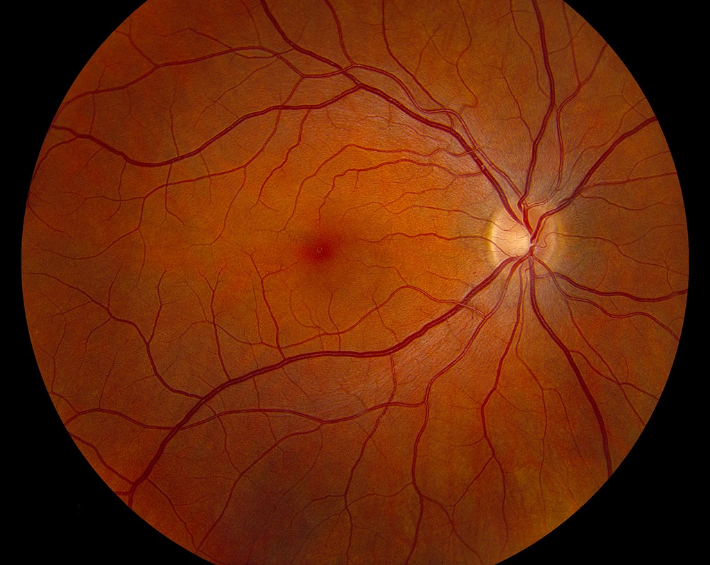
A normal fundus has a pink-orange background and a sharply outlined optic disc.
Four vessel arcades leave the disc and curve around the macula.
The macula looks slightly darker, with a bright foveal reflex in young eyes.
Examining the Normal Fundus
First, view the posterior pole with a 78D or 90D lens at the slit lamp.
Then, sweep the periphery to the ora serrata with indirect ophthalmoscopy.
In addition, baseline color or widefield photos make later comparison easier.
Fundus Explorer Pro
Photograph the retinal findings described here with the phone already in your pocket — 22 D optics and built-in illumination in one handheld unit.
From Choroida — the team behind this siteThe Optic Disc
The healthy disc is slightly vertically oval, roughly 1.5 to 2 mm across.
Its rim looks orange-pink because perfused nerve fibers fill it.
The nasal margin often looks less sharp than the temporal margin.
Also, a temporal crescent of pigment or bare sclera is common.
The Optic Cup and Neuroretinal Rim
The cup is the central pale depression without axons.
Because cup size follows disc size, large discs carry large cups.
Many eyes follow the ISNT pattern, with the inferior rim thickest.
However, many normal eyes break this rule (see cup-to-disc ratio pitfalls).
Retinal Vessels in the Normal Fundus
Arteries look lighter red and narrower than veins, at a width ratio near 2 to 3.
They also carry a thin central light reflex.
At crossings, a healthy artery passes the vein without nicking or deflection.
Spontaneous venous pulsation on the disc is common in healthy eyes, while its absence alone proves little.
The Macula and Fovea
The fovea lies about two disc diameters temporal to the disc, slightly below its center.
Luteal pigment and denser RPE pigment make this zone look darker.
As a result, the fovea may appear as a subtle dark spot.
Also, the foveal center is free of visible vessels.
Foveal Reflex in a Normal Fundus
The foveal reflex is a pinpoint of light from the concave foveal pit.
Young eyes may also show a ring-shaped reflex around the macula.
Both reflexes fade with age, so their absence in older eyes is expected.
However, a missing reflex in a young eye warrants OCT to look for edema.
Normal Fundus Variants by Ethnicity
Fundus color depends on pigment in the RPE and choroid.
Patients of African descent often show a darker, slate-gray, sometimes granular background.
East Asian eyes often show a more uniform brown-orange tone.
In contrast, lightly pigmented eyes look orange-red, with visible choroidal vessels (see tessellated fundus).
Therefore, judge symmetry between eyes before suspecting a dystrophy.
Age Changes in the Normal Fundus
Aging dulls the reflexes and reduces the nerve fiber layer sheen.
Arteries may narrow slightly, and their light reflex can broaden.
A few small drusen under 63 micrometers count as normal aging changes.
However, medium or large drusen place the eye in an AMD category.
The Young Fundus Sheen
Children show a bright, glistening reflex along the arcades from the internal limiting membrane.
Scattered sparkling points are also normal (see Gunn dots).
As a result, this sheen should not be mistaken for exudate.
Common Disc Variants
Several benign variants appear in otherwise healthy eyes.
- Myelinated nerve fibers: white feathery patches that hide vessels
- Tilted disc: oblique nerve entry, common in myopia
- Peripapillary atrophy: a pale crescent beside the disc
- Cilioretinal artery: a vessel emerging from the temporal rim
- Bergmeister papilla: a glial remnant on the disc surface
The Periphery of a Normal Fundus
The peripheral retina looks thinner, so the choroid shows through more.
Vortex vein ampullae appear as dark choroidal whorls near the equator.
Long posterior ciliary nerves run horizontally at 3 and 9 o’clock.
In addition, paving stone and peripheral cystoid degeneration are common benign findings in adults.
Normal Fundus on OCT
OCT shows a smooth foveal pit and continuous outer retinal bands.
Inner retinal layers thin out toward the foveal center.
The choroid also thins with age and axial myopia.
Therefore, compare thickness values with age-matched normative data.
Counseling Patients About a Normal Fundus
Patients often ask whether a feature on their photo means disease.
Explain that disc size, pigment and sheen differ between people, much like skin tone.
Then, show both eyes side by side when symmetry supports a benign finding.
Finally, arrange follow-up when a variant overlaps with glaucoma or early AMD.
Pitfalls to Avoid
- Do not call a large cup glaucoma without judging disc size
- Never label dark, mottled pigment as dystrophy without comparing eyes
- Do not read the young retinal sheen as exudate
- Avoid treating small hard drusen as AMD


Document what you see
Two smartphone imaging tools built for everyday clinic use — one for the slit lamp, one for the fundus.
From Choroida — the team behind this siteReferences
- Ferris FL 3rd, Wilkinson CP, Bird A, et al. Clinical classification of age-related macular degeneration. Ophthalmology. 2013;120:844-851.
- Jonas JB, Budde WM, Panda-Jonas S. Ophthalmoscopic evaluation of the optic nerve head. Surv Ophthalmol. 1999;43:293-320.
- Harizman N, Oliveira C, Chiang A, et al. The ISNT rule and differentiation of normal from glaucomatous eyes. Arch Ophthalmol. 2006;124:1579-1583.
- Jacks AS, Miller NR. Spontaneous retinal venous pulsation: aetiology and significance. J Neurol Neurosurg Psychiatry. 2003;74:7-9.
- Bowling B. Kanski’s Clinical Ophthalmology: A Systematic Approach. 8th ed. Elsevier; 2016.
- American Academy of Ophthalmology. Basic and Clinical Science Course, Section 12: Retina and Vitreous. San Francisco: AAO; 2023.

Writes and reviews the clinical articles on Educate. Every post is checked against peer-reviewed sources before it goes live.