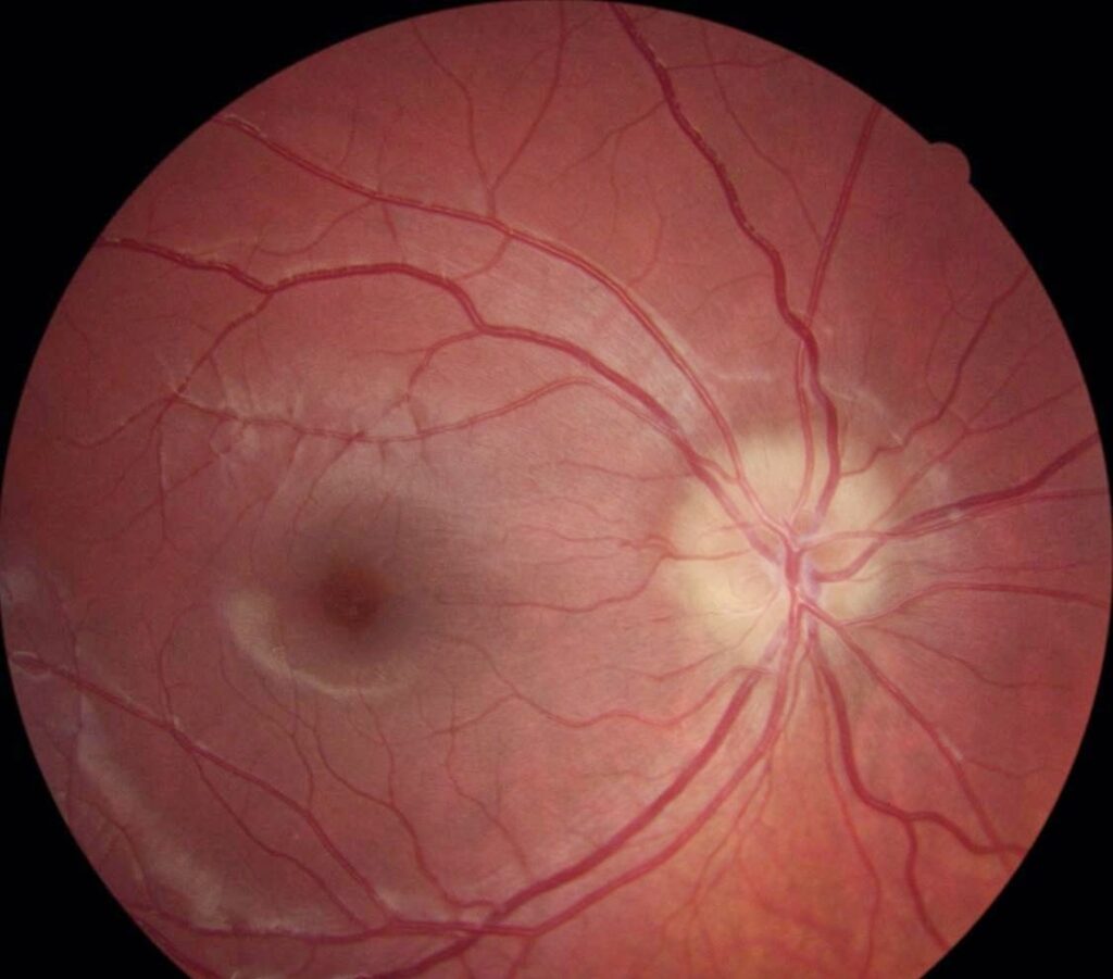
Crowded optic disc describes a small optic nerve head with little or no visible cup.
Many clinicians call it a “disc at risk” because the configuration predisposes to ischemic optic neuropathy.
However, most eyes with this anatomy never develop disease.
What a Crowded Optic Disc Is
The neuroretinal rim fills almost the entire surface of a small disc.
As a result, the cup is tiny or absent, and the margins may look slightly indistinct.
The appearance is a normal anatomical variant.
It gains clinical weight when an examiner mistakes it for swelling or when ischemia strikes.
Anatomy of the Small Scleral Canal
Roughly a million ganglion cell axons pass through the scleral canal.
In a small canal, the same axons occupy less space and pile up at the disc surface.
Therefore, no room remains for a central cup.
Jonas and colleagues showed that disc area varies widely in normal eyes and that cup size follows disc size.
Fundus Explorer Pro
Photograph the retinal findings described here with the phone already in your pocket — 22 D optics and built-in illumination in one handheld unit.
From Choroida — the team behind this siteWho Has a Crowded Optic Disc
Hyperopic eyes often have small globes and small scleral canals.
Patients with optic disc drusen also tend to have small canals.
In addition, the fellow eye of a patient with ischemic optic neuropathy usually shows the same configuration.
Many healthy emmetropes have it too, so the finding carries no diagnosis by itself.
Examination Signs of a Crowded Optic Disc
A 78 D or 90 D lens at the slit lamp gives the stereoscopic view you need.
The disc looks small, full and slightly raised above the retinal plane.
Vessels emerge centrally and may branch early or in an anomalous pattern.
However, they stay clearly visible as they cross the disc margin.
The color stays pink without hyperemia, while the peripapillary nerve fiber layer looks normal.
Crowded Optic Disc Versus True Swelling
True edema obscures segments of major vessels at the disc margin.
It also brings venous engorgement, peripapillary hemorrhages, cotton wool spots or retinal folds.
In contrast, a crowded disc lacks these signs and stays stable over time.
When doubt remains, compare old photographs and follow a structured workup (see optic disc swelling causes).
Separating Buried Drusen
Buried drusen produce a lumpy elevated disc that can mimic edema.
Autofluorescence highlights superficial drusen, while B-scan ultrasound detects calcified ones.
Next, enhanced depth OCT shows deeper drusen as signal-poor bodies with a hyperreflective margin.
The consortium recommendations define these OCT features in detail (see optic disc drusen).
OCT in the Crowded Optic Disc
Peripapillary nerve fiber layer thickness is usually normal or only mildly increased.
Marked thickening with inward bowing of peripapillary Bruch membrane favors raised intracranial pressure in the right context.
Also, optic nerve head scans show a small Bruch membrane opening, which confirms the small canal.
Therefore, a baseline scan gives an objective reference for later visits.
Why the Label “Disc at Risk” Persists
The phrase came from studies of fellow eyes in nonarteritic ischemic optic neuropathy.
Beck and colleagues found smaller cup-to-disc ratios in affected patients than in controls.
Burde then described the crowded disc as a structural risk factor.
Hayreh, however, has argued that anatomy alone cannot explain the event and that vascular factors are required.
Crowded Optic Disc as an Ischemic Risk Factor
One theory holds that axons in a tight canal have little room to swell.
Then, a modest drop in perfusion may trigger axoplasmic stasis and edema.
As a result, swelling could compress capillaries further and create a compartment-like cycle.
This mechanism remains a hypothesis, and its exact sequence is still debated.
Factors That Add Risk
- Systemic hypertension and diabetes
- Nocturnal hypotension, for example from evening antihypertensive dosing
- Obstructive sleep apnea
- Optic disc drusen in the same eye
- Drugs reported in association, such as amiodarone and phosphodiesterase-5 inhibitors
Glaucoma Assessment in a Crowded Optic Disc
A small disc with a small cup can still hide glaucomatous damage.
Because rim area is limited, modest loss changes the appearance very little.
Therefore, judge the rim against disc size and look for notching or a disc hemorrhage (see disc hemorrhage in glaucoma).
Counseling and Follow-Up
Explain that the disc shape is inborn and usually harmless.
Avoid the word “risk” without context, because patients may hear it as a coming event.
Next, address modifiable factors such as blood pressure, sleep apnea and glucose control.
Finally, record baseline photographs and advise urgent review for any sudden painless visual loss.
Pitfalls to Avoid
- Do not call a crowded disc papilledema without vessel obscuration or other swelling signs
- Never skip drusen imaging when the disc looks lumpy
- Avoid reading a small cup as proof that glaucoma is absent


Document what you see
Two smartphone imaging tools built for everyday clinic use — one for the slit lamp, one for the fundus.
From Choroida — the team behind this siteReferences
- Beck RW, Servais GE, Hayreh SS. Anterior ischemic optic neuropathy. IX. Cup-to-disc ratio and its role in pathogenesis. Ophthalmology. 1987;94:1503-1508.
- Burde RM. Optic disk risk factors for nonarteritic anterior ischemic optic neuropathy. Am J Ophthalmol. 1993;116:759-764.
- Hayreh SS. Ischemic optic neuropathy. Prog Retin Eye Res. 2009;28:34-62.
- Jonas JB, Gusek GC, Naumann GO. Optic disc, cup and neuroretinal rim size, configuration and correlations in normal eyes. Invest Ophthalmol Vis Sci. 1988;29:1151-1158.
- Malmqvist L, Bursztyn L, Costello F, et al. The Optic Disc Drusen Studies Consortium recommendations for diagnosis of optic disc drusen using optical coherence tomography. J Neuroophthalmol. 2018;38:299-307.

Writes and reviews the clinical articles on Educate. Every post is checked against peer-reviewed sources before it goes live.